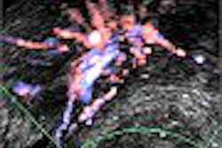
Wall compliance and stiffness measurement may be useful in evaluating the success of endovascular repair for abdominal aortic aneurysms (AAA), according to a study by U.K. researchers.
The ultrasound study, done in 57 patients to assess aortic wall compliance as an indicator for rupture risk in AAA, also concluded that endovascular repair (EVR) leaves patients with greater wall stiffness than those undergoing open surgical repair.
"Postrepair compliance is influenced by actual wall stiffness, intraluminal or intrasac pressure, pulse pressure, and also the type of stent (balloon or self-expandable) and the effect of the thrombus," said Dr. Amine Sekhri, who presented the study findings at the 2004 RSNA conference in Chicago. Sekhri's group is from the medical imaging and endovascular units of The Middlesex Hospital and University College London Hospitals in London.
The study consisted of 29 subjects in the prerepair group and 32 in the postrepair group. Eight patients were in both groups. The mean patient age was 78 years. Of the 32 patients in the postrepair group, 26 underwent endovascular repair and six had open surgical repair. There were endoleaks or endotension in eight patients with EVR. The rest had no complications.
Ultrasound was done using a HDI 5000 scanner (Philips Medical Systems, Andover, MA). The cine-loop images were analyzed offsite to measure the aortic diameter during systole and diastole using wall-tracking software, HDILab (ATL, Bothwell, WA).
Brachial blood pressure was measured, and elastic modulus (Ep) and stiffness (β) calculated. Ep and β were calculated at the neck of the aneurysm, inflection points, and midsac levels. "Patients with endoleaks had significantly elevated Ep and ß compared with those without endoleaks," the researchers stated.
According to previous research, Ep and β have an inverse relationship with aortic wall compliance. This allows a better estimate of AAA rupture risk than taking aneurysm size alone into consideration (European Journal of Vascular Endovascular Surgery, June 1998, Vol. 15:6; pp. 472-477).
Based on the preoperative and postoperative Ep and β results from the current study, Sekhri also observed that the compliance measurement agreed with wall stress distribution in AAA. And that maximum stress is not necessarily located in the maximum diameter.
Other studies to assess the risk of rupture have found wall stress to be distributed with distinct regions of high and low stress. Knowledge of this stress distribution will be a better indicator than going only by the diameter of the aneurysm (Journal of Vascular Surgery, April 2000, Vol. 31:4, pp. 760-769).
Aortic wall compliance measurement in AAA can detect the potential prerepair state of AAA and evaluate the success of EVR, with the advantage of being noninvasive and low-cost with ultrasound, Sekhri concluded.
By N. Shivapriya
AuntMinnie.com contributing writer
March 15, 2005
Related Reading
MRI for carotid artery wall thickness measures up to US gold standard, February 3, 2005
Older men should be screened for aortic aneurysm, February 1, 2005
New treatment for aortic aneurysm triggers debate, October 15, 2004
Copyright © 2005 AuntMinnie.com


![Examples of ultrasound findings and techniques. (A) Images in a 39-year-old male patient with a mass in the left thigh. The mass is heterogeneous on the B-mode US image (compared with the patient in D) and showed increased microvascularity (superb microvascular imaging [SMI]) and shear-wave elastography (SWE) values. Undifferentiated pleomorphic sarcoma was diagnosed at biopsy (with pleomorphic rhabdomyosarcoma in surgical specimen). (B) Images in an 18-year-old male patient with a mass in the left leg. The mass is hypoechoic on the B-mode image, with no other findings suggestive of malignancy. The lesion is in contact with the cortex of the tibia, which is slightly irregular. CT revealed a doubtful anteromedial tibial erosion. The microvascular study demonstrated high vascularization, suggestive of malignancy. Periosteal Ewing sarcoma was diagnosed with both histologic and immunohistochemical confirmation. (C) Images in a 69-year-old female patient with a lump growing on the outside of the left leg. Multiple SWE examinations were performed (please note the high values obtained in the measurements, whereas the color map highlights the stiffness relative to adjacent tissues). SMI showed areas of increased vascularization to target for sampling. Undifferentiated spindle cell sarcoma was diagnosed at biopsy, with residual leiomyosarcoma in the surgical specimen after neoadjuvant therapy. (D) Images in a 56-year-old female patient with a mass in the right thigh. The mass is heterogeneous at both B-mode ultrasound (similar to patient A) and MRI (coronal T2-weighted spectral attenuated inversion recovery [SPAIR]; T1-weighted pre-contrast and postcontrast imaging), which even shows uptake after the administration of paramagnetic contrast material, which is traditionally suggestive of malignancy. Low values at SMI and elastography are suggestive of benignity. Spindle cell lipoma was diagnosed at biopsy, with atypical spindle cell lipomatous tumor in the surgical specimen.](https://img.auntminnie.com/mindful/smg/workspaces/default/uploads/2026/08/images-radiol250278fig2.APCFLSvX6p.jpg?auto=format%2Ccompress&fit=crop&h=100&q=70&w=100)


![Examples of ultrasound findings and techniques. (A) Images in a 39-year-old male patient with a mass in the left thigh. The mass is heterogeneous on the B-mode US image (compared with the patient in D) and showed increased microvascularity (superb microvascular imaging [SMI]) and shear-wave elastography (SWE) values. Undifferentiated pleomorphic sarcoma was diagnosed at biopsy (with pleomorphic rhabdomyosarcoma in surgical specimen). (B) Images in an 18-year-old male patient with a mass in the left leg. The mass is hypoechoic on the B-mode image, with no other findings suggestive of malignancy. The lesion is in contact with the cortex of the tibia, which is slightly irregular. CT revealed a doubtful anteromedial tibial erosion. The microvascular study demonstrated high vascularization, suggestive of malignancy. Periosteal Ewing sarcoma was diagnosed with both histologic and immunohistochemical confirmation. (C) Images in a 69-year-old female patient with a lump growing on the outside of the left leg. Multiple SWE examinations were performed (please note the high values obtained in the measurements, whereas the color map highlights the stiffness relative to adjacent tissues). SMI showed areas of increased vascularization to target for sampling. Undifferentiated spindle cell sarcoma was diagnosed at biopsy, with residual leiomyosarcoma in the surgical specimen after neoadjuvant therapy. (D) Images in a 56-year-old female patient with a mass in the right thigh. The mass is heterogeneous at both B-mode ultrasound (similar to patient A) and MRI (coronal T2-weighted spectral attenuated inversion recovery [SPAIR]; T1-weighted pre-contrast and postcontrast imaging), which even shows uptake after the administration of paramagnetic contrast material, which is traditionally suggestive of malignancy. Low values at SMI and elastography are suggestive of benignity. Spindle cell lipoma was diagnosed at biopsy, with atypical spindle cell lipomatous tumor in the surgical specimen.](https://img.auntminnie.com/mindful/smg/workspaces/default/uploads/2026/08/images-radiol250278fig2.APCFLSvX6p.jpg?auto=format%2Ccompress&fit=crop&h=112&q=70&w=112)