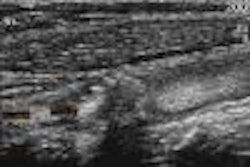
(Ultrasound Review) The purpose of this study was to characterize the sonographic appearances of galactoceles. This is the most common benign breast lesion in lactating patients. Ultrasound is the modality of choice for diagnosis, because women of childbearing age tend to have breasts of the increased density that are difficult to assess mammographically. Ultrasound also may be used to guide needle aspiration. The Journal of Clinical Ultrasound published this study earlier this year.
A retrospective review by radiologists from the All India Institute of Medical Sciences in New Delhi, found 10 patients had been diagnosed with galactoceles over a five-year period. Analysis of results revealed that "at the time of their examination, all patients had been lactating (2-9 months postpartum) and had had a unilateral, painless, palpable lump, not associated with fever," they reported.
All patients underwent ultrasound of both breasts using a high-resolution linear array transducer. Typical ultrasound characteristics included a mean diameter of 3.6 cm; well defined, thin echogenic walls; homogeneous (60%) appearance; heterogeneous with anechoic fluid rim (40%); and posterior enhancement (90%).
Eight patients had a single lesion, one had two, and one had 12 lesions. Ultrasound-guided needle aspiration was performed on the lesions and a white milk product containing fat and debris was withdrawn. According to the authors, "needle aspiration alone was therapeutic in eight patients." Of the remaining patients, one had an infected galactocele, and the other had multiple lesions that required surgical removal.
Galactoceles are easily differentiated from simple cysts, but not atypical cysts that may in fact be infected cysts, abscesses, intraductal papilloma, and intracystic carcinoma. The debris contained in galactoceles is similar in appearance to a mural nodule with debris. The authors recommend that color Doppler imaging should be used to demonstrate flow in solid components, and confirm or rule-out a malignancy. Occasionally, galactoceles show ultrasound characteristics typical of fibroadenoma and carcinoma.
They concluded that since there are overlapping findings, the most suitable means of diagnosing galactoceles was through fine-needle aspiration. This had the added benefit of removing the problem in the majority of cases, they said.
"Sonographic appearances of galactoceles"Sawhney, S. et al
Department of radiodiagnosis, All India Institute of Medical Sciences, New Delhi, India
Journal of Clinical Ultrasound 2002 January; 30:18-22
By Ultrasound Review
April 9, 2002
Copyright © 2002 AuntMinnie.com
![Examples of ultrasound findings and techniques. (A) Images in a 39-year-old male patient with a mass in the left thigh. The mass is heterogeneous on the B-mode US image (compared with the patient in D) and showed increased microvascularity (superb microvascular imaging [SMI]) and shear-wave elastography (SWE) values. Undifferentiated pleomorphic sarcoma was diagnosed at biopsy (with pleomorphic rhabdomyosarcoma in surgical specimen). (B) Images in an 18-year-old male patient with a mass in the left leg. The mass is hypoechoic on the B-mode image, with no other findings suggestive of malignancy. The lesion is in contact with the cortex of the tibia, which is slightly irregular. CT revealed a doubtful anteromedial tibial erosion. The microvascular study demonstrated high vascularization, suggestive of malignancy. Periosteal Ewing sarcoma was diagnosed with both histologic and immunohistochemical confirmation. (C) Images in a 69-year-old female patient with a lump growing on the outside of the left leg. Multiple SWE examinations were performed (please note the high values obtained in the measurements, whereas the color map highlights the stiffness relative to adjacent tissues). SMI showed areas of increased vascularization to target for sampling. Undifferentiated spindle cell sarcoma was diagnosed at biopsy, with residual leiomyosarcoma in the surgical specimen after neoadjuvant therapy. (D) Images in a 56-year-old female patient with a mass in the right thigh. The mass is heterogeneous at both B-mode ultrasound (similar to patient A) and MRI (coronal T2-weighted spectral attenuated inversion recovery [SPAIR]; T1-weighted pre-contrast and postcontrast imaging), which even shows uptake after the administration of paramagnetic contrast material, which is traditionally suggestive of malignancy. Low values at SMI and elastography are suggestive of benignity. Spindle cell lipoma was diagnosed at biopsy, with atypical spindle cell lipomatous tumor in the surgical specimen.](https://img.auntminnie.com/mindful/smg/workspaces/default/uploads/2026/08/images-radiol250278fig2.APCFLSvX6p.jpg?auto=format%2Ccompress&fit=crop&h=100&q=70&w=100)


![Examples of ultrasound findings and techniques. (A) Images in a 39-year-old male patient with a mass in the left thigh. The mass is heterogeneous on the B-mode US image (compared with the patient in D) and showed increased microvascularity (superb microvascular imaging [SMI]) and shear-wave elastography (SWE) values. Undifferentiated pleomorphic sarcoma was diagnosed at biopsy (with pleomorphic rhabdomyosarcoma in surgical specimen). (B) Images in an 18-year-old male patient with a mass in the left leg. The mass is hypoechoic on the B-mode image, with no other findings suggestive of malignancy. The lesion is in contact with the cortex of the tibia, which is slightly irregular. CT revealed a doubtful anteromedial tibial erosion. The microvascular study demonstrated high vascularization, suggestive of malignancy. Periosteal Ewing sarcoma was diagnosed with both histologic and immunohistochemical confirmation. (C) Images in a 69-year-old female patient with a lump growing on the outside of the left leg. Multiple SWE examinations were performed (please note the high values obtained in the measurements, whereas the color map highlights the stiffness relative to adjacent tissues). SMI showed areas of increased vascularization to target for sampling. Undifferentiated spindle cell sarcoma was diagnosed at biopsy, with residual leiomyosarcoma in the surgical specimen after neoadjuvant therapy. (D) Images in a 56-year-old female patient with a mass in the right thigh. The mass is heterogeneous at both B-mode ultrasound (similar to patient A) and MRI (coronal T2-weighted spectral attenuated inversion recovery [SPAIR]; T1-weighted pre-contrast and postcontrast imaging), which even shows uptake after the administration of paramagnetic contrast material, which is traditionally suggestive of malignancy. Low values at SMI and elastography are suggestive of benignity. Spindle cell lipoma was diagnosed at biopsy, with atypical spindle cell lipomatous tumor in the surgical specimen.](https://img.auntminnie.com/mindful/smg/workspaces/default/uploads/2026/08/images-radiol250278fig2.APCFLSvX6p.jpg?auto=format%2Ccompress&fit=crop&h=112&q=70&w=112)