For detecting prostate cancer, 3-D power Doppler ultrasound adds diagnostic heft to its 2-D counterpart, according to a Russian study presented at the 2002 European Congress of Radiology in Vienna. The technique may also improve the targeting of prostate biopsies.
"It's well known that prostate cancer growth is associated with tumoral angiogenesis and the presence of neovascularization," said Dr. Marina Kisliakova from the postgraduate research and education center of Moscow's Outpatient Clinic No. 1. "Three-dimensional power Doppler (can) visualize the whole prostate vessel map and reveal tumoral vessels. However, the potential value of this method ... compared with power Doppler is insufficiently studied."
The study examined 97 patients, ages 52-85 (average 63.3 ± 4.1 years) with suspected prostate cancer based on the results of digital rectal examination and prostate-specific antigen (PSA) test levels.
Each patient underwent three successive ultrasound exams, including transrectal ultrasound (TRUS) using B-mode, power Doppler, and 3-D power Doppler, all on an HDI 5000 scanner (Philips Medical Systems). The radiologists were aware of the results of each prior exam, which could have affected the results to a small degree, Kisliakova told AuntMinnie.com.
B-mode data were acquired using map 3, medium persistence, penetration option, and maximum frame rate. Power Doppler and 3-D power Doppler images were acquired using map 3, medium wall filter, PRF 700 Hz, and the resistance-flow option.
The B-mode examination assessed prostate echogenicity and volume, as well as the presence of focal lesions and their location, echogenicity, and structure. The power Doppler and 3-D power Doppler exams also examined the vascularity of focal lesions, blood-flow patterns (hypervascularity), and the presence of chaotic vascularity and deformed vessels, Kisliakova said.
"All vascularity characteristics were assessed comparing with the contralateral prostate side," she said.
Kisliakova, along with colleagues Dr. Veronika Gajonova and Dr. Alexander Zubarev, compared the power Doppler results with the 3-D power Doppler data. All patients underwent targeted US-guided biopsies, and random sextant US-guided biopsies were performed in patients who did not have palpable or US-visible lesions.
The ultrasound results were then compared with histopathology, which revealed 63 cases of prostate cancer (64.9%), 14 cases of benign prostate hypertrophy (BPH, 14.4%), 14 cases of chronic prostatitis (14.4%), and 6 cases of the precancerous condition known as prostate intraepithelial neoplasia (PIN, 6.3%).
According to the results, B-mode TRUS found 68 focal lesions, which resulted in 33 cancer diagnoses. B-mode plus power Doppler found an additional 29 lesions (n=62) and yielded an additional 14 cancer diagnoses (n=47). The addition of 3-D power Doppler to the two previous modes added the visualization of 17 focal lesions (n=114) and yielded 16 additional histologically proven cancer diagnoses (n=63).
The addition of 3-D power Doppler to 2-D also yielded significantly more cases of focal hypervascularity and deformed vessels, according to the chart below. Kisliakova said that while only a few vessels could be seen using 2-D power Doppler, 3-D provided a complete vascular picture that was often diagnostic by itself. Some false-positive cases were eliminated when a lack of abnormal vasculature was noted on 3-D power Doppler.
 |
Positive and negative predictive values were 66% and 52%, respectively, for B-mode TRUS; 81% and 75%, respectively, for B-mode plus power Doppler; and 87% and 82%, respectively, for B-mode plus power Doppler plus 3-D power Doppler.
Sensitivity and specificity were 75% and 55%, respectively, for B-mode TRUS; 80% and 77%, respectively, for B-mode plus power Doppler; and reached 90% and 86%, respectively, for B-mode plus power Doppler plus 3-D power Doppler, Kisliakova said.
 |
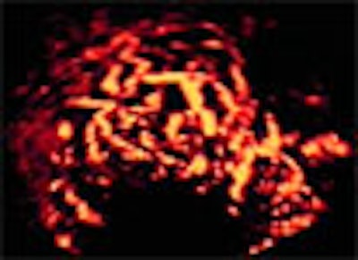
| In images of a prostate cancer patient above, 3-D power Doppler US image (bottom) details diffuse hypervascular zone with deformed asymmetric vessels in the right lobe. Vascular abnormalities are not visualized in B-mode TRUS image (top left) and are only suggested in power Doppler image (top right). |
 |
| Three-dimensional power Doppler US also helped diagnose a patient with prostate intraepithelial neoplasia (PIN), above, showing neovascular abnormalities in a hypoechoic lesion in the right peripheral zone. (Top left image is B-mode TRUS, top right is B-mode close-up, lower left is 3-D power Doppler, and lower right is 3-D power Doppler close-up.) Images and graphics courtesy of Dr. Marina Kisliakova. |
Adding 3-D power Doppler allowed the visualization of low-flow tumoral vessels, and assessment of the degree of vascularity in focal lesions as compared to the normal contralateral side, Kisliakova concluded. Thus, the researchers were able to define blood-flow patterns and neovascularity features, and detect additional lesions with changed vascularity characteristics.
"The 3-D power Doppler TRUS examination is more informative than power Doppler TRUS in the detection of prostate cancer," she said. "(It) can be used as a diagnostic tool in addition to the complex examination of patients with suspected prostate cancer, and may be useful for more accurate (targeting of) biopsies."
By Eric BarnesAuntMinnie.com staff writer
March 25, 2002
Related Reading
Criss-crossing vascularity reliably predicts testicular cancer, March 7, 2002
Ultrasound positions prostate cancer patients for therapy, November 7, 2001
Copyright © 2002 AuntMinnie.com