B-flow Doppler ultrasound may soon replace duplex Doppler in the evaluation of carotid stenosis. The B-flow technique images blood reflectors directly, providing a real-time picture of flow in a display that resembles an angiogram. A group of researchers from the University of Vermont College of Medicine and the Fletcher Allen Health Care facility in Burlington, VT, recently tested the capabilities of B-flow ultrasound in measuring stenosis severity.
Dr. Brian Garra presented the results of his team’s research at the 2002 American Institute of Ultrasound in Medicine meeting in Nashville last month. Garra said that duplex Doppler classifies stenosis only into broad ranges of 20% or more. The group sought to determine if B-flow ultrasound could provide greater accuracy and precision in stenosis measurement than current Doppler methods.
B-flow technology utilizes digitally encoded ultrasound to decode and display signals. The data is then enhanced and equalized to separate tissue and blood signals to provide an image of simultaneous tissue and flow. The image also displays plaque margins that are not obscured by overlay, and its spatial resolution is preserved, Garra said.
The researchers performed gray-scale and B-flow ultrasound on 45 carotid arteries in 36 patients, who also underwent either contrast arteriography (8 patients) or MR angiography (MRA). According to Garra, 5 of the arterial studies were excluded because MRAs were read as uncertain in 4 patients, and because the B-flow exam couldn't be read in 1 patient due to problems with the acquisition technique.
The ultrasound exams were performed on a GE Medical Systems (Waukesha, WI) Logiq 700 Expert super-premium scanner equipped with a 7-MHz linear-array transducer (LA 739 probe). Both static images and video loops of the studies were saved and then viewed on an eFilm Medical (Toronto) workstation, version 1.53.
 |
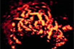
| A B-flow image showing shadowing plaque and jet. Image courtesy of Dr. Brian Garra. |
The team then measured the luminal diameter and stenosis using the longitudinal and cross-sectional video of the carotids at the mid common carotid artery, distal common carotid artery, proximal internal carotid artery, and the mid-to-distal internal carotid artery. Stenosis percentage values for ultrasound, arteriography, and MRA were calculated by the researchers using the lumen diameter measured more distally as the "normal" reference.
Garra and his team found that the B-flow stenosis estimate was within (±) 5% of the angiographic value for 18 carotids, and within (±) 10% for the remaining 22 carotids. They also reported that the average study quality was higher for longitudinal images than for transverse images. In addition, the average difference in stenosis percentage across all carotids, as compared to MRA, was lower in the longitudinal images.
 |
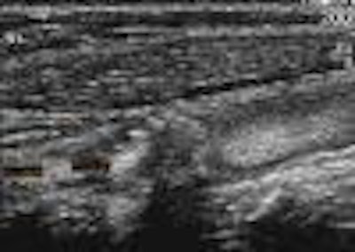
| The researchers measured stenosis on both static and video-loop B-flow images. Image courtesy of Dr. Brian Garra. |
Although the technology shows great promise, Garra cautioned that a few issues need to be resolved before it will see widespread clinical use. On his wish list of technological improvements is a means of representing 3-D blood flow, as well as an automated boundary detection tool. He said the most important factor in achieving quality B-flow results is the acquisition and interpretation of the images themselves.
"On a carotid duplex examination, you just put the cursor in the vessel in a couple of places and you look for the highest velocity," Garra said. "On a B-flow you’re actually going to have to measure down to a fraction of a millimeter. A really narrow stenosis is going to look like a thread on the screen. If you think about it, if it’s a 1-mm lumen and you’re off by a millimeter, you’re off by 50%."
By Jonathan S. BatchelorAuntMinnie.com staff writer
April 4, 2002
Related Reading
Color carotid ultrasonography findings helps pinpoint stroke risk, December 24, 2001
Carotid endarterectomy with normal findings: Is there need for early duplex scan?, November 6, 2001
Ultrasound inadequate as sole means of determining need for carotid intervention, October 15, 2001
Transcranial Doppler an insensitive measure of cerebrovascular reserve, August 24, 2001
Duplex criteria for determination of 50% or greater carotid stenosis, May 23, 2001
Copyright © 2002 AuntMinnie.com