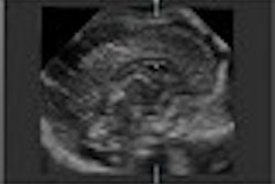
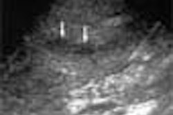
(Ultrasound Review) Radiologists in Heidelberg, Germany recently evaluated the use of contrast-enhanced harmonic imaging in diagnosing vesicoureteral reflux in pediatric patients. The aim of the study was to compare contrast harmonic imaging, tissue harmonic imaging, and conventional B-mode imaging, "with regard to sonomorphology and detection and ease of demonstration of reflux, if present."
Previous studies have proven that voiding urosonography with Levovist (Schering, Berlin, Germany) has a higher sensitivity and specificity than radiological reflux studies.
Fifty-four children with indications including urinary tract infection, follow-up after conservative or surgical management, and pyelectasis and megaureter were included in the study. Initial conventional sonography was followed by introduction of a contrast agent, Levovist, into the bladder through the urethra.
"The contrast-enhanced sonography was conducted by scanning the bladder and each kidney in transverse and longitudinal planes, from ventral and dorsal views, consecutively in B-mode using fundamental, contrast harmonic, and tissue harmonic imaging modalities," the authors reported.
Results showed that tissue harmonic imaging was the best modality for demonstration of both the retrovesical space and the renal pelvis. Forty-one percent of children displayed reflux, and the refluxing microbubbles were better demonstrated using the harmonic modes compared with conventional ultrasound.
Harmonic imaging detected reflux in eight children that appeared normal with conventional ultrasound after introduction of contrast. Comparing contrast and tissue harmonic modalities in reflux detection, there was no significant difference, but the authors felt that contrast harmonics was more suitable when only a few microbubbles were shown in the renal pelvis. The anatomy of the retrovesical space and renal pelvis was best visualized using tissue harmonic imaging.
According to the authors, using harmonic imaging affords improved diagnostic sensitivity, and reductions in ultrasound contrast amount and subsequent cost, and learning curve and procedure duration.
"Visualization of the urinary tract and detection of ultrasound contrast media is significantly improved by the use of the harmonic imaging modalities. When both fundamental and harmonic imaging options are available, we recommend harmonic imaging for contrast-enhanced sonographic diagnosis of vesicoureteral reflux," the authors concluded.
"Contrast-enhanced harmonic imaging for the diagnosis of vesicoureteral reflux in pediatric patients"
Kassa Daarge et al
Dept of Pediatric Radiology, Radiological Clinic, Ruprecht-Karls University Heidelberg, Im Neuenheimer Feld 153, 69120 Heidelberg, Germany
AJR 2001 (December); 177:1411–1415
By Ultrasound Review
February 14, 2002
Copyright © 2002 AuntMinnie.com

![Examples of ultrasound findings and techniques. (A) Images in a 39-year-old male patient with a mass in the left thigh. The mass is heterogeneous on the B-mode US image (compared with the patient in D) and showed increased microvascularity (superb microvascular imaging [SMI]) and shear-wave elastography (SWE) values. Undifferentiated pleomorphic sarcoma was diagnosed at biopsy (with pleomorphic rhabdomyosarcoma in surgical specimen). (B) Images in an 18-year-old male patient with a mass in the left leg. The mass is hypoechoic on the B-mode image, with no other findings suggestive of malignancy. The lesion is in contact with the cortex of the tibia, which is slightly irregular. CT revealed a doubtful anteromedial tibial erosion. The microvascular study demonstrated high vascularization, suggestive of malignancy. Periosteal Ewing sarcoma was diagnosed with both histologic and immunohistochemical confirmation. (C) Images in a 69-year-old female patient with a lump growing on the outside of the left leg. Multiple SWE examinations were performed (please note the high values obtained in the measurements, whereas the color map highlights the stiffness relative to adjacent tissues). SMI showed areas of increased vascularization to target for sampling. Undifferentiated spindle cell sarcoma was diagnosed at biopsy, with residual leiomyosarcoma in the surgical specimen after neoadjuvant therapy. (D) Images in a 56-year-old female patient with a mass in the right thigh. The mass is heterogeneous at both B-mode ultrasound (similar to patient A) and MRI (coronal T2-weighted spectral attenuated inversion recovery [SPAIR]; T1-weighted pre-contrast and postcontrast imaging), which even shows uptake after the administration of paramagnetic contrast material, which is traditionally suggestive of malignancy. Low values at SMI and elastography are suggestive of benignity. Spindle cell lipoma was diagnosed at biopsy, with atypical spindle cell lipomatous tumor in the surgical specimen.](https://img.auntminnie.com/mindful/smg/workspaces/default/uploads/2026/08/images-radiol250278fig2.APCFLSvX6p.jpg?auto=format%2Ccompress&fit=crop&h=100&q=70&w=100)


![Examples of ultrasound findings and techniques. (A) Images in a 39-year-old male patient with a mass in the left thigh. The mass is heterogeneous on the B-mode US image (compared with the patient in D) and showed increased microvascularity (superb microvascular imaging [SMI]) and shear-wave elastography (SWE) values. Undifferentiated pleomorphic sarcoma was diagnosed at biopsy (with pleomorphic rhabdomyosarcoma in surgical specimen). (B) Images in an 18-year-old male patient with a mass in the left leg. The mass is hypoechoic on the B-mode image, with no other findings suggestive of malignancy. The lesion is in contact with the cortex of the tibia, which is slightly irregular. CT revealed a doubtful anteromedial tibial erosion. The microvascular study demonstrated high vascularization, suggestive of malignancy. Periosteal Ewing sarcoma was diagnosed with both histologic and immunohistochemical confirmation. (C) Images in a 69-year-old female patient with a lump growing on the outside of the left leg. Multiple SWE examinations were performed (please note the high values obtained in the measurements, whereas the color map highlights the stiffness relative to adjacent tissues). SMI showed areas of increased vascularization to target for sampling. Undifferentiated spindle cell sarcoma was diagnosed at biopsy, with residual leiomyosarcoma in the surgical specimen after neoadjuvant therapy. (D) Images in a 56-year-old female patient with a mass in the right thigh. The mass is heterogeneous at both B-mode ultrasound (similar to patient A) and MRI (coronal T2-weighted spectral attenuated inversion recovery [SPAIR]; T1-weighted pre-contrast and postcontrast imaging), which even shows uptake after the administration of paramagnetic contrast material, which is traditionally suggestive of malignancy. Low values at SMI and elastography are suggestive of benignity. Spindle cell lipoma was diagnosed at biopsy, with atypical spindle cell lipomatous tumor in the surgical specimen.](https://img.auntminnie.com/mindful/smg/workspaces/default/uploads/2026/08/images-radiol250278fig2.APCFLSvX6p.jpg?auto=format%2Ccompress&fit=crop&h=112&q=70&w=112)