
Oxford Brain Diagnostics highlighted a study showing that its Cortical Disarray Measurement (CDM) technology corresponded with the distribution of autopsy-confirmed pathology in patients with frontotemporal lobar degeneration (FTLD), the disease process underlying frontotemporal dementia (FTD).
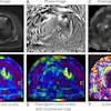
Researchers compared diffusion MRI measurements taken during life with postmortem findings across 14 cortical regions in 36 autopsy-confirmed cases of behavioral variant FTD: 19 with FTLD-tau and 17 with FTLD-TDP. They used PerpPD+, a CDM-derived measure of cortical microstructure, to assess whether the MRI signal tracked pathology in the outer versus deeper cortical layers.
CDM patterns were associated with how pathology was distributed across cortical layers, and the relationship differed between the two molecular forms, according to the company. It was strongest in FTLD-TDP, where the MRI measurement followed the higher concentration of pathology in the outer layers. The findings help establish an important connection between what can be measured noninvasively in the living brain and the microscopic pathology ultimately observed at autopsy, Oxford Brain Diagnostics said.
The study is being presented at the 15th International Conference on Frontotemporal Dementias and was conducted at the University of Pennsylvania, led David Irwin, MD.


.fFmgij6Hin.png?auto=compress%2Cformat&fit=crop&h=100&q=70&w=100)

.fFmgij6Hin.png?auto=compress%2Cformat&fit=crop&h=167&q=70&w=250)


