3D slice-to-volume MRI can help accurately describe and measure the fetal optic pathway, according to findings presented April 16 at the American Roentgen Ray Society (ARRS) annual meeting in Hawaii.
The research, presented via a scientific online poster and led by Eric Juang from Creighton University School of Medicine's Phoenix Regional Campus, found that this MRI method successfully visualized more fetal optic pathways than unprocessed fetal MRI.
"Our preliminary results ... demonstrate the promises and utility of this technique," Juang said in a statement from the ARRS.
The fetal optic pathway is important because disrupted development can lead to permanent visual impairment. This makes early detection important, but the target area is tiny at about a millimeter or less.
The researchers noted that ultrasound-based techniques have limited fetal presentation and low reproducibility. Additionally, they pointed out that conventional MRI is inconsistent in visualizing fetal optic presentation due to low resolution.
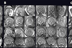
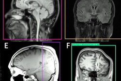
3D slice-to-volume MRI, however, requires six or more T2 single shot fast spin-echo sequences with multiple planes. After semiautomated tools are used to segment the images, 3D images are then reconstructed by intensity matching of the segmentations.
Juang and colleagues wanted to describe this technique to visualize the fetal optic pathway, as well as compare the percentage of times the pathway is visualized between this method and conventional MRI. Ultimately, the team wants to create a nomogram based on their findings.
The researchers looked at retrospective data from 70 fetal MRI scans collected between 2020 and 2022. They found that fetal optic pathway was visualized in 55 cases via 3D slice-to-volume MRI. Conventional unprocessed fetal MRI meanwhile visualized just nine cases.
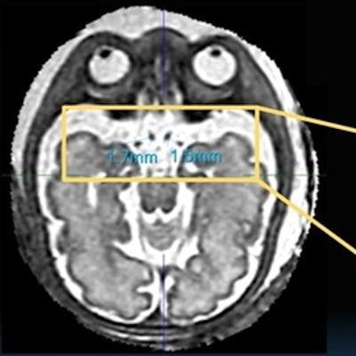
 Measurements complete by medical student under supervision of two pediatric neuroradiologists with more than eight years of reading experience. Image and caption courtesy of the ARRS.
Additionally, among the 3D slice-to-volume cases, prechiasmatic optic nerve width was successfully measured bilaterally in 53 cases, optic chiasm width in all cases, and bilateral optic tract width in 30 cases.
The team also used a linear regression fit to estimate the relationship between optic chiasm (OCW) width in millimeters in normal fetuses and gestational age (GA) in weeks. It came up with the equation of OCW = 0.11 × GA + 2.0 (R^2 = 0.30). In other words, about 30% of optic chiasm and nerve width can be attributed to gestational age. Similarly, the relationship between prechiasmatic (PC) optic nerve width and GA was estimated as PC = 0.04 × GA + 0.24 (R^2 = 0.34).
However, Juang and colleagues also noted that the 3D method is less accessible than ultrasound and requires more images for reconstruction.
Juang said further results are pending but added that the improved resolution seen through the 3D method makes way for improved precision in fetal optic pathway measurements. Advantages he noted for 3D slice-to-volume MRI include being less susceptible to motion artifacts and being unaffected by fetal position.
The poster presentation received the online poster summa cum laude award for ARRS 2023.