
MRI is illuminating what may be a new form of dementia in World Trade Center (WTC) first responders with both cognitive impairment and post-traumatic stress disorder (PTSD) in a study published August 11 in the Journal of Alzheimer's Disease.
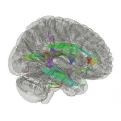
A team from Stony Brook University in New York found that a 3D MRI imaging technique called diffusion tractography imaged the white brain matter of 99 first responders to the attack on the World Trade Center in 2001. It determined that the white matter of those study participants with PTSD and cognitive impairment looked different than that of first responders with cognitive impairment but no PTSD. In particular, first responders with both cognitive impairment and PTSD had cerebellar atrophy, a manifestation of white matter degradation that appears to be new among symptoms of dementia.
"Overall, the study supports the view that responders with CI have neurological changes consistent with neurodegenerative disease, but they are inconclusive as to the type of disease," said lead author Sean Clouston, PhD, in a statement released by the university on August 22. "Our findings do show that dementia due to PTSD is clearly different from non-PTSD dementia in this responder population."


![Overview of the study design. (A) The fully automated deep learning framework was developed to estimate body composition (BC) (defined as subcutaneous adipose tissue [SAT] in liters; visceral adipose tissue [VAT] in liters; skeletal muscle [SM] in liters; SM fat fraction [SMFF] as a percentage; and intramuscular adipose tissue [IMAT] in deciliters) from MRI. The fully automated framework comprised one model (model 1) to quantify different BC measures (SAT, VAT, SM, SMFF, and IMAT) as three-dimensional (3D) measures from whole-body MRI scans. The second model (model 2) was trained to identify standardized anatomic landmarks along the craniocaudal body axis (z coordinate field), which allowed for subdividing the whole-body measures into different subregions typically examined on clinical routine MRI scans (chest, abdomen, and pelvis). (B) BC was quantified from whole-body MRI in over 66,000 individuals from two large population-based cohort studies, the UK Biobank (UKB) (36,317 individuals) and the German National Cohort (NAKO) (30,291 individuals). Bar graphs show age distribution by sex and cohort. BMI = body mass index. (C) After the performance assessment of the fully automated framework, the change in BC measures, distributions, and profiles across age decades were investigated. Age-, sex-, and height-adjusted body composition reference curves were calculated and made publicly available in a web-based z-score calculator (https://circ-ml.github.io).](https://img.auntminnie.com/mindful/smg/workspaces/default/uploads/2026/05/body-comp.XgAjTfPj1W.jpg?auto=format%2Ccompress&fit=crop&h=112&q=70&w=112)

