Brain MRI helps predict postsurgical outcomes in children who have sustained traumatic brain injury (TBI) and have undergone surgery for the condition, according to a study published January 21 in the Journal of Neurosurgery: Pediatrics.
The findings could help clinicians better counsel families of pediatric TBI patients, wrote a team led by Dr. Cordell Baker, a neurosurgeon at the University of Utah in Salt Lake City.
"Development of outcome predictors in these patients is necessary to ensure accurate family counseling and to appropriately guide postoperative management," the group wrote.
Traumatic brain injury is the most common cause of death and disability among children around the world, with more than 600,000 emergency room visits in the U.S. each year for these injuries, the group reported, citing data from the U.S. Centers for Disease Control and Prevention (CDC).
Children presenting in the emergency department with TBI often undergo surgery to relieve pressure on the brain caused by internal bleeding, and while most recover satisfactorily, some do not. It can be difficult to predict which children with TBI will have poorer results after surgery.
Previous studies exploring the use of MRI for predicting outcomes in children with TBI before surgery have found a link between diffuse axonal injury-type lesions (i.e., the tearing of the brain's long connecting nerve fibers) and worse postsurgical outcomes. But more research is needed to determine whether MRI would be useful for predicting outcomes after these children have already undergone surgery.
"No study to date has evaluated MRI for prognostication in children who have undergone either craniectomy or craniotomy after severe traumatic brain injury," Baker's team wrote. "The establishment of an accurate outcome predictor in these patients is necessary to guide appropriate clinical management and accurately counsel family members on prognosis and expectations when these injuries occur."
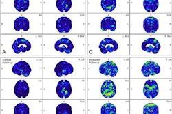
To address this knowledge gap, the group conducted a study that included 92 children who underwent craniectomy or craniotomy for TBI between 2010 and 2019. Of these, 43 received a postoperative brain MRI exam within four months of surgery. The exams used susceptibility-weighted imaging (SWI) and fluid-attenuated inversion recovery (FLAIR) sequences to identify any cerebral lesions (both bleeding and nonbleeding) related to diffuse axonal injury; lesions were categorized as superficial, deep, or brainstem.
The investigators tracked the following variables for any association with poor outcome:
- The location of the diffuse axonal injury
- Glasgow Coma Scale score
- Pediatric trauma score
- Cause of injury
- Time to surgery
They reported outcome results using the King's Outcome Scale for Childhood Head Injury (KOSCHI), which is a five-point, pediatric version of the Glasgow Coma Scale (1 = death, 2 = vegetative state, 3 = severe disability, 4 = moderate disability, and 5 = good, without any effect on functioning).
Of 43 children with TBI who underwent postsurgical MRI (median day, 5), median Glasgow Coma Scale score on admission to the emergency department was 4 and the most prevalent pediatric trauma score was 0 to 5, or life-threatening (53%). The most common cause of the brain injury was falls (33%), and the most common hemorrhage type was subdural hematoma (60%).
The postsurgery brain MRI exams identified two statistically significant variables to be associated with poor outcomes postsurgery: cerebral herniation (p = 0.027) and the location of the diffuse axonal injury, specifically in the brainstem (p = 0.037).
The authors noted that 84% of children in this subset of patients with postsurgery MRI exams went on to recover, but they cautioned that it's important to keep in mind the variables that may indicate poor postsurgical outcomes. Of particular concern is the fact that the group found that 53% of children in the study sample who sustained severe brain injuries and underwent surgery for them never received follow-up MRI.
"Our results indicate that postoperative MRI in this population may be especially important for prognostication," Baker and colleagues noted. "However, in our initial patient identification, we found that many children (53%) who presented with severe traumatic brain injury and underwent surgery never received a postoperative brain MRI."
The study suggests that more research on the use of MRI postsurgery in children who have sustained TBI is needed, according to the authors.
"Our results revealed that diffuse axonal injury-type lesions in the brainstem and evidence of cerebral herniation may indicate a poorer prognosis; however, more studies with larger cohorts are needed to make definitive conclusions," they concluded.