Prerecorded, available throughout the meeting | SPR-NR-38
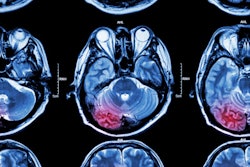
Brain MRI with an ultrashort echo time protocol is a cost-effective way of classifying mild or unspecific neurologic symptoms compared to standard- or reduced-dose CT, according to research presented in this prerecorded talk.The findings could have both cost and patient care benefits, as patients could be treated prophylactically for subsequent strokes, presenter Dr. Daniel Puhr-Westerheide of Ludwig Maximilian University of Munich and colleagues noted in the study abstract.
For the study, Puhr-Westerheide's team explored the cost-effectiveness of short-protocol brain MRI after negative noncontrast CT for identifying minor strokes in emergency department patients with mild or unspecific neurological symptoms. The group used a Markov model to assess the cost-effectiveness of this strategy, with willingness to pay set at $100,000 per quality-adjusted life year (QALY); the model was based on a simulated follow-up time frame of 30 years.
The investigators found that using short-protocol MRI in this manner lowered costs from $27,109 for a protocol that consisted of CT only to $26,304 and boosted patients' quality of life years from 14.25 in the CT-only group to 14.31 in the short-protocol MRI group.
"Additional short-protocol MRI in emergency patients with mild and unspecific neurological symptoms enables timely secondary prophylaxis through detection of minor strokes, resulting in lower costs and higher cumulative QALYs," Puhr-Westerheide and colleagues concluded.


![Overview of the study design. (A) The fully automated deep learning framework was developed to estimate body composition (BC) (defined as subcutaneous adipose tissue [SAT] in liters; visceral adipose tissue [VAT] in liters; skeletal muscle [SM] in liters; SM fat fraction [SMFF] as a percentage; and intramuscular adipose tissue [IMAT] in deciliters) from MRI. The fully automated framework comprised one model (model 1) to quantify different BC measures (SAT, VAT, SM, SMFF, and IMAT) as three-dimensional (3D) measures from whole-body MRI scans. The second model (model 2) was trained to identify standardized anatomic landmarks along the craniocaudal body axis (z coordinate field), which allowed for subdividing the whole-body measures into different subregions typically examined on clinical routine MRI scans (chest, abdomen, and pelvis). (B) BC was quantified from whole-body MRI in over 66,000 individuals from two large population-based cohort studies, the UK Biobank (UKB) (36,317 individuals) and the German National Cohort (NAKO) (30,291 individuals). Bar graphs show age distribution by sex and cohort. BMI = body mass index. (C) After the performance assessment of the fully automated framework, the change in BC measures, distributions, and profiles across age decades were investigated. Age-, sex-, and height-adjusted body composition reference curves were calculated and made publicly available in a web-based z-score calculator (https://circ-ml.github.io).](https://img.auntminnie.com/mindful/smg/workspaces/default/uploads/2026/05/body-comp.XgAjTfPj1W.jpg?auto=format%2Ccompress&fit=crop&h=100&q=70&w=100)


![Overview of the study design. (A) The fully automated deep learning framework was developed to estimate body composition (BC) (defined as subcutaneous adipose tissue [SAT] in liters; visceral adipose tissue [VAT] in liters; skeletal muscle [SM] in liters; SM fat fraction [SMFF] as a percentage; and intramuscular adipose tissue [IMAT] in deciliters) from MRI. The fully automated framework comprised one model (model 1) to quantify different BC measures (SAT, VAT, SM, SMFF, and IMAT) as three-dimensional (3D) measures from whole-body MRI scans. The second model (model 2) was trained to identify standardized anatomic landmarks along the craniocaudal body axis (z coordinate field), which allowed for subdividing the whole-body measures into different subregions typically examined on clinical routine MRI scans (chest, abdomen, and pelvis). (B) BC was quantified from whole-body MRI in over 66,000 individuals from two large population-based cohort studies, the UK Biobank (UKB) (36,317 individuals) and the German National Cohort (NAKO) (30,291 individuals). Bar graphs show age distribution by sex and cohort. BMI = body mass index. (C) After the performance assessment of the fully automated framework, the change in BC measures, distributions, and profiles across age decades were investigated. Age-, sex-, and height-adjusted body composition reference curves were calculated and made publicly available in a web-based z-score calculator (https://circ-ml.github.io).](https://img.auntminnie.com/mindful/smg/workspaces/default/uploads/2026/05/body-comp.XgAjTfPj1W.jpg?auto=format%2Ccompress&fit=crop&h=112&q=70&w=112)


