Use of a portable low-field MRI scanner is feasible for neuroimaging patients in the intensive care unit (ICU), according to a study published September 8 in JAMA Neurology. The research results suggest that there could be a way to make neuroimaging more accessible for critically ill patients.
"For ICUs, access to MRI is limited, and the risks of transporting patients with critical illness are well documented," wrote a team led by Dr. Kevin Sheth of Yale University School of Medicine in New Haven. "Risks to inpatient populations and clinicians are potentially increased when considering infection control issues, as illustrated by the COVID-19 pandemic. This report helps fill an important gap in the topic of obtaining neuroimaging for patients with critical illness and potential neuropathology."
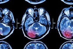
Imaging is a key way to evaluate brain injury, and MRI is often the first-line imaging modality for this indication, the group noted. But MRI exams are conducted in controlled environments to manage the high-strength magnetic fields the technology produces, making access to MRI difficult for patients in intensive care.
"The traditional neuroimaging workflow requires patient transport to dedicated hospital imaging suites," the investigators wrote. "This operational paradigm has been necessary to ensure patient safety in and around high-field scanners but has rendered MRI largely inaccessible in the setting of critical illness."
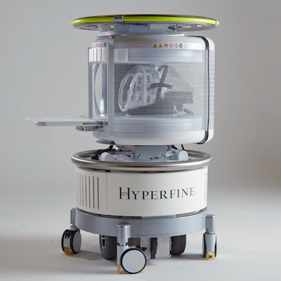
Sheth's team included founders and cofounders of Hyperfine Research, which developed a low-field MRI scanner (0.064 tesla) that can produce images that offer clinically useful data outside of the MRI suite -- even in the presence of ferromagnetic items at bedside. The scanner does not use cryogens and is powered by a standard power outlet. The U.S. Food and Drug Administration (FDA) cleared the latest generation of the device in August; this study is the first clinical report regarding its performance, the authors wrote.
The study consisted of 50 patients admitted to Yale New Haven Hospital ICU for either neurological injury or COVID-19 between October 2019 and May 2020. Patients presented with the following conditions:
- Ischemic stroke (9)
- Hemorrhagic stroke (12)
- Subarachnoid hemorrhage (2)
- Traumatic brain injury (3)
- Brain tumor (4)
- COVID-19 with altered mental status (20)
The study participants underwent portable MRI exams at a median of five days after admission to the ICU. MRI sequences included T1-weighted, T2-weighted, T2 fluid-attenuated inversion recovery, and diffusion-weighted, according to Sheth and colleagues. The scanner's head coil was positioned over the patient at bedside, and the exam did not impede ICU equipment.
The portable scanner identified neurological findings in 97% patients who did not have COVID-19 and in 40% of those who did, the team found. No complications or adverse events resulted from use of the scanner. Twenty-nine of 30 patients without COVID-19 and 11 of 20 patients with COVID-19 also underwent CT and conventional MR imaging, and all the portable MRI findings were consistent with reports from these exams.
The point-of-care MRI scanner could be a valuable tool for the care of critically ill patients, according to the team.
"[Our] experience demonstrates that low-field, portable MRI can be deployed successfully into intensive care settings," the authors concluded. "This approach may hold promise for portable assessment of neurological injury in other scenarios, including the emergency department, mobile stroke units, and resource-limited environments."