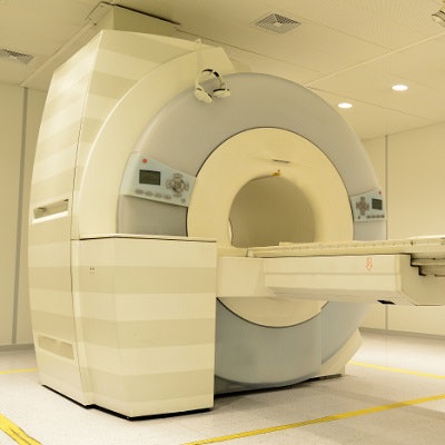
Optimizing costs with beneficial patient outcomes is a constant goal in healthcare. In that regard, the use of MR angiography (MRA) to screen and follow up patients who may have intracranial aneurysms linked to kidney disease can work well from both cost and clinical perspectives, according to a study published online February 19 in Radiology.
Researchers analyzed several clinical scenarios for patients with autosomal dominant polycystic kidney disease (ADPKD) to detect and manage aneurysms. They found that MRA screening every five years is cost-effective for patients with ADPKD. If an aneurysm is detected, patients should receive annual surveillance MRA and should be converted to treatment if the aneurysm continues growing.
"Our study results indicate that MR angiography screening of patients with ADPKD every five years, and annual MR angiography follow-up in patients in whom intracranial aneurysm is detected, is the optimal strategy," wrote the researchers led by Dr. Ajay Malhotra from the department of radiology and biomedical imaging at the Yale School of Medicine. "It remains so when the life expectancy is more than six years."
ADPKD affects between 1 in 400 and 1 in 1,000 people and is associated with major extrarenal complications. Intracranial aneurysms occur in approximately 10% of ADPKD patients, and among them, 23% have multiple aneurysms. Individuals with the disease are also more susceptible to aneurysm rupture, at an average age of 41 years -- some 10 years earlier than the general population.
The question is whether patients with ADPKD and intracranial aneurysms should be screened before the aneurysms have a chance to rupture. MRA is one possible tool, but the benefits of screening are unknown due to "the lack of data from randomized, prospective clinical trials," Malhotra and colleagues noted.
To better determine the potential utility of MRA as a screening tool in this population, the researchers took clinical parameters from recently published studies with large cohorts or meta-analyses specific to patients with ADPKD. They then crafted a decision-analytics model to assess different strategies for ADPKD patients with intracranial aneurysms:
- No MRA screening
- A one-time MRA screening with an annual MRA follow-up
- MRA screening every five years with endovascular treatment
- MRA screening every five years with annual MRA follow-up
- MRA screening every five years with biennial MRA follow-up
The researchers then computed clinical outcomes and costs with each scenario. They assessed outcomes in terms of quality-adjusted life years (QALYs) and net monetary benefit.
Their calculations determined that MRA screening every five years with annual follow-up was the best strategy with a QALY of 25.86, compared with a QALY of 25.84 for MRA screening every five years with biennial MRA follow-up (the difference in QALY of 0.04 translates to two weeks of life in perfect health, the researchers noted).
| Cost-effectiveness of MRA screening strategies for intracranial aneurysms in ADPKD patients | ||
| Strategy | Cost | QALY |
| One-time MRA screening with annual follow-up | $16,978 | 25.82 |
| MRA screening every 5 years with biennial MRA follow-up | $19,485 | 25.84 |
| MRA screening every 5 years with MRA annual follow-up | $19,839 | 25.86 |
| MRA screening every 5 years with endovascular treatment | $84,593 | 25.35 |
| No MRA screening | $147,919 | 22.99 |
As for which approach might yield the greatest health benefit, the researchers conducted 10,000 MRA simulations based on the presence of an intracranial aneurysm. MRA screening every five years with annual MRA follow-up was superior to one-time MRA screening with annual MRA follow-up in 7,126 (71%) of the iterations.