
A second imaging facility is reporting problems with Apple devices that appear to be related to the operation of the center's MRI scanner. Nearly 10 late-model Apple iPhones and Watches were permanently disabled at a Delaware center after it ramped down its MRI magnet.
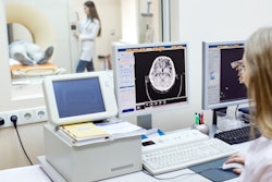
CNMRI is an imaging facility in Dover, DE, that specializes in neurology and sleep medicine. It operates a 1.5-tesla MRI magnet and also performs studies such as polysomnography, nerve conduction, and home sleep studies, according to Dr. Robert Varipapa, a neurologist at the center.
In mid-October, field service engineers from an imaging OEM arrived and ramped the magnet down and then back up again. Immediately thereafter, staff members at the center who owned Apple devices with wireless charging reported that their devices were disabled. Approximately eight or nine devices were affected, according to Varipapa.
Only newer-model Apple products such as the iPhone 8 and iPhone 10 were affected, he added. Those with older models didn't experience any problems, nor did staff with Android phones.
The Delaware center's experience is similar to that of an Illinois hospital that also reported conflicts between its MRI scanner and iPhones. That site reported that nearly 40 iPhones stopped working after the installation of a new MRI scanner. The problem was attributed to helium gas that may have leaked during the installation and found its way into the mechanical workings of the phones.
But there are also crucial differences between the Illinois incident and the experience at the Delaware center. For one thing, the Delaware site never experienced a helium leak, to Varipapa's knowledge. Also, while the Illinois site reported problems with Apple models at the iPhone 6 level and above, in Delaware the problem was restricted to newer models with wireless charging -- no iPhone 6 devices were affected, Varipapa told AuntMinnie.com.
Finally, at the Illinois hospital, some of the iPhones began working again after the helium inside the devices apparently dissipated. At CNMRI, all of the smartphones were permanently disabled, and staff had to get new ones.
Varipapa said CNMRI's physicist told him that the center's experience is not an uncommon one. The physicist has heard that some field service engineers tell staff members to place smartphones in their cars' glove boxes when MRI magnets are being serviced, he said.


![Overview of the study design. (A) The fully automated deep learning framework was developed to estimate body composition (BC) (defined as subcutaneous adipose tissue [SAT] in liters; visceral adipose tissue [VAT] in liters; skeletal muscle [SM] in liters; SM fat fraction [SMFF] as a percentage; and intramuscular adipose tissue [IMAT] in deciliters) from MRI. The fully automated framework comprised one model (model 1) to quantify different BC measures (SAT, VAT, SM, SMFF, and IMAT) as three-dimensional (3D) measures from whole-body MRI scans. The second model (model 2) was trained to identify standardized anatomic landmarks along the craniocaudal body axis (z coordinate field), which allowed for subdividing the whole-body measures into different subregions typically examined on clinical routine MRI scans (chest, abdomen, and pelvis). (B) BC was quantified from whole-body MRI in over 66,000 individuals from two large population-based cohort studies, the UK Biobank (UKB) (36,317 individuals) and the German National Cohort (NAKO) (30,291 individuals). Bar graphs show age distribution by sex and cohort. BMI = body mass index. (C) After the performance assessment of the fully automated framework, the change in BC measures, distributions, and profiles across age decades were investigated. Age-, sex-, and height-adjusted body composition reference curves were calculated and made publicly available in a web-based z-score calculator (https://circ-ml.github.io).](https://img.auntminnie.com/mindful/smg/workspaces/default/uploads/2026/05/body-comp.XgAjTfPj1W.jpg?auto=format%2Ccompress&fit=crop&h=100&q=70&w=100)


![Overview of the study design. (A) The fully automated deep learning framework was developed to estimate body composition (BC) (defined as subcutaneous adipose tissue [SAT] in liters; visceral adipose tissue [VAT] in liters; skeletal muscle [SM] in liters; SM fat fraction [SMFF] as a percentage; and intramuscular adipose tissue [IMAT] in deciliters) from MRI. The fully automated framework comprised one model (model 1) to quantify different BC measures (SAT, VAT, SM, SMFF, and IMAT) as three-dimensional (3D) measures from whole-body MRI scans. The second model (model 2) was trained to identify standardized anatomic landmarks along the craniocaudal body axis (z coordinate field), which allowed for subdividing the whole-body measures into different subregions typically examined on clinical routine MRI scans (chest, abdomen, and pelvis). (B) BC was quantified from whole-body MRI in over 66,000 individuals from two large population-based cohort studies, the UK Biobank (UKB) (36,317 individuals) and the German National Cohort (NAKO) (30,291 individuals). Bar graphs show age distribution by sex and cohort. BMI = body mass index. (C) After the performance assessment of the fully automated framework, the change in BC measures, distributions, and profiles across age decades were investigated. Age-, sex-, and height-adjusted body composition reference curves were calculated and made publicly available in a web-based z-score calculator (https://circ-ml.github.io).](https://img.auntminnie.com/mindful/smg/workspaces/default/uploads/2026/05/body-comp.XgAjTfPj1W.jpg?auto=format%2Ccompress&fit=crop&h=112&q=70&w=112)


