
Researchers at Cedars-Sinai are giving the term multitasking a new meaning by developing an MRI technique that eliminates breath-holds and shortens cardiac scan time, according to a paper published online April 9 in Nature Biomedical Engineering.
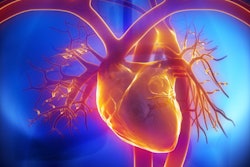
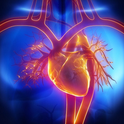
The MR multitasking approach creates what they call 6D images of a beating heart and blood flow with diagnostic accuracy and enhanced patient comfort.
Currently, patients undergoing cardiac MRI scans are asked to hold their breath while images are being acquired. The process is particularly troublesome and unreliable for patients with irregular heartbeats or breathing problems.
"It is challenging to obtain good cardiac magnetic resonance images because the heart is beating incessantly, and the patient is breathing, so the motion makes the test vulnerable to errors," said Dr. Shlomo Melmed, dean of the Cedars-Sinai medical faculty, in a statement from Cedars-Sinai Medical Center. "By novel approaches to this longstanding problem, this research team has found a unique solution to improve cardiac care for patients around the world for years to come."
The researchers tested the MR multitasking technique on 10 healthy volunteers and 10 cardiac patients, continuously acquiring image data with no breath-holds. They then separated overlapping sources of motion into multiple time dimensions after the scan was completed.
MR multitasking was found to be accurate and more comfortable for patients, said Debiao Li, PhD, the study's senior author. In addition, the investigators were able to complete three cardiac MRI tests in as little as 90 seconds.
"Now we collect all the data throughout the entire test and sort it afterwards," Li said. "We get full control after the test, as opposed to trying to control the body's natural movement during imaging."
By incorporating motion and time into the MR multitasking process, the researchers are able to create images with six dimensions.
"Our videos are 6D because we can play them back four different ways," said lead author Anthony Christodoulou, PhD, a research scientist at Cedars-Sinai. "We can playback cardiac motion, respiratory motion, and two different tissue processes that reveal cardiac health."
MR multitasking is currently being used at several medical centers in the U.S. and China. The group hopes to expand the technique's use to other diseases.


![Overview of the study design. (A) The fully automated deep learning framework was developed to estimate body composition (BC) (defined as subcutaneous adipose tissue [SAT] in liters; visceral adipose tissue [VAT] in liters; skeletal muscle [SM] in liters; SM fat fraction [SMFF] as a percentage; and intramuscular adipose tissue [IMAT] in deciliters) from MRI. The fully automated framework comprised one model (model 1) to quantify different BC measures (SAT, VAT, SM, SMFF, and IMAT) as three-dimensional (3D) measures from whole-body MRI scans. The second model (model 2) was trained to identify standardized anatomic landmarks along the craniocaudal body axis (z coordinate field), which allowed for subdividing the whole-body measures into different subregions typically examined on clinical routine MRI scans (chest, abdomen, and pelvis). (B) BC was quantified from whole-body MRI in over 66,000 individuals from two large population-based cohort studies, the UK Biobank (UKB) (36,317 individuals) and the German National Cohort (NAKO) (30,291 individuals). Bar graphs show age distribution by sex and cohort. BMI = body mass index. (C) After the performance assessment of the fully automated framework, the change in BC measures, distributions, and profiles across age decades were investigated. Age-, sex-, and height-adjusted body composition reference curves were calculated and made publicly available in a web-based z-score calculator (https://circ-ml.github.io).](https://img.auntminnie.com/mindful/smg/workspaces/default/uploads/2026/05/body-comp.XgAjTfPj1W.jpg?auto=format%2Ccompress&fit=crop&h=100&q=70&w=100)


![Overview of the study design. (A) The fully automated deep learning framework was developed to estimate body composition (BC) (defined as subcutaneous adipose tissue [SAT] in liters; visceral adipose tissue [VAT] in liters; skeletal muscle [SM] in liters; SM fat fraction [SMFF] as a percentage; and intramuscular adipose tissue [IMAT] in deciliters) from MRI. The fully automated framework comprised one model (model 1) to quantify different BC measures (SAT, VAT, SM, SMFF, and IMAT) as three-dimensional (3D) measures from whole-body MRI scans. The second model (model 2) was trained to identify standardized anatomic landmarks along the craniocaudal body axis (z coordinate field), which allowed for subdividing the whole-body measures into different subregions typically examined on clinical routine MRI scans (chest, abdomen, and pelvis). (B) BC was quantified from whole-body MRI in over 66,000 individuals from two large population-based cohort studies, the UK Biobank (UKB) (36,317 individuals) and the German National Cohort (NAKO) (30,291 individuals). Bar graphs show age distribution by sex and cohort. BMI = body mass index. (C) After the performance assessment of the fully automated framework, the change in BC measures, distributions, and profiles across age decades were investigated. Age-, sex-, and height-adjusted body composition reference curves were calculated and made publicly available in a web-based z-score calculator (https://circ-ml.github.io).](https://img.auntminnie.com/mindful/smg/workspaces/default/uploads/2026/05/body-comp.XgAjTfPj1W.jpg?auto=format%2Ccompress&fit=crop&h=112&q=70&w=112)


