
Real-time intraoperative MRI (RT-IMRI) could be a valuable tool to guide and monitor the successful transplantation of stem cells used in therapy for Parkinson's disease, according to a study published online September 14 in Cell Transplantation.
Researchers from the University of Wisconsin-Madison are using RT-IMRI to transplant neurons derived from induced pluripotent stem cells (iPSCs) into the brains of nonhuman primates modeled with Parkinson's disease. So far, the imaging technique has provided enhanced visualization and monitoring of the procedure and helps cell survival.
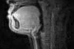
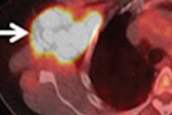
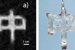
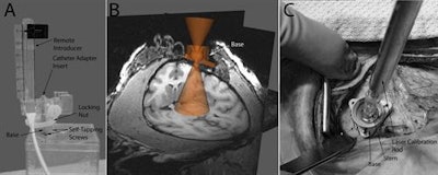
Images show technology and methodology involved in MRI-guided transplantation of neural stem cells into the Parkinsonian brain. Courtesy of Cell Transplantation.Lead author Dr. Marina Emborg, PhD, and colleagues developed an MRI-compatible trajectory guidance system that has been successful for intraoperative MRI. Most recently, they upgraded the system for real-time targeting and guidance, which allows for real-time pressure readings that can prevent clogging during cell delivery.
Upon postmortem brain analysis, the researchers found that transplanted cells grafted and survived well in the test animals after transplantation.


![Overview of the study design. (A) The fully automated deep learning framework was developed to estimate body composition (BC) (defined as subcutaneous adipose tissue [SAT] in liters; visceral adipose tissue [VAT] in liters; skeletal muscle [SM] in liters; SM fat fraction [SMFF] as a percentage; and intramuscular adipose tissue [IMAT] in deciliters) from MRI. The fully automated framework comprised one model (model 1) to quantify different BC measures (SAT, VAT, SM, SMFF, and IMAT) as three-dimensional (3D) measures from whole-body MRI scans. The second model (model 2) was trained to identify standardized anatomic landmarks along the craniocaudal body axis (z coordinate field), which allowed for subdividing the whole-body measures into different subregions typically examined on clinical routine MRI scans (chest, abdomen, and pelvis). (B) BC was quantified from whole-body MRI in over 66,000 individuals from two large population-based cohort studies, the UK Biobank (UKB) (36,317 individuals) and the German National Cohort (NAKO) (30,291 individuals). Bar graphs show age distribution by sex and cohort. BMI = body mass index. (C) After the performance assessment of the fully automated framework, the change in BC measures, distributions, and profiles across age decades were investigated. Age-, sex-, and height-adjusted body composition reference curves were calculated and made publicly available in a web-based z-score calculator (https://circ-ml.github.io).](https://img.auntminnie.com/mindful/smg/workspaces/default/uploads/2026/05/body-comp.XgAjTfPj1W.jpg?auto=format%2Ccompress&fit=crop&h=112&q=70&w=112)


