Is the patient a psychopath, a lifelong substance abuser, or both? The question is a critical one for victims and the criminal justice system alike. According to a study published online Monday in the Archives of General Psychiatry, MRI is beginning to shine a light on differences in the brains of violent offenders with and without substance abuse problems, which could help reveal causes and potential cures for those who commit violent offenses.
The German study found that gray-matter volumes in certain brain regions of violent offenders are different from those in individuals who don't commit violent acts. Long-term substance use is also associated with different gray-matter alterations that can be distinguished from those of nonabusing violent offenders.
Specifically, larger gray-matter volumes in the mesolimbic reward system may be associated with violent acts, and reduced gray-matter volumes in the frontal cortex and premotor areas characterize men with histories of substance use, according to researchers from University of Duisburg-Essen and several other academic centers in Germany and the U.K.
"Our hypothesis that men with [substance use disorders (SUDs], compared with those without SUDs, would be characterized by alterations in the prefrontal cortex was confirmed," wrote Boris Schiffer, PhD, and colleagues (Arch Gen Psychiatry, June 6, 2011).
Violent behavior causes much suffering and is a costly social problem, accounting for 6.5% and 11.9% of the gross domestic products of Germany and the U.S., respectively, the authors noted. "Consequently, furthering our understanding of the small group of men who commit most of the violent crimes is a priority," they wrote.
Previous studies have pointed to differences in gray-matter volume at MRI in these troubled populations. Unfortunately, their design didn't account for long histories of substance use, resulting in possible incorrect interpretations; the authors of the present study aimed to address by this by identifying alterations in gray-matter volume associated with violent behavior separately from those associated with SUDs.
The research group recruited 51 men, including 24 violent offenders (mean 3.3 violent crimes), from penitentiaries, psychiatric hospitals, psychiatric outpatient hospitals, and communities in Germany.
The 24 offenders were divided into two groups: 12 with a lifetime history of SUDs and 12 without such a designation. Similarly, the nonoffender group included 13 men with SUDs and 14 healthy men with no criminal histories. The four groups were matched by age and education level.
All participants underwent 1.5-tesla MRI scans (Sonata, Siemens Healthcare) using a 3D T1-weighted rapid-acquisition gradient-echo sequence acquiring 160 contiguous 10-mm sagittal slices (240 x 240 mm2 field-of-view, 5º flip angle, and a voxel size of 1.0 x 0.9 x 1.0 mm). Data were processed using Matlab 7.4 (MathWorks) and statistical parametric mapping software. Voxel-based morphometry was used to assess gray-matter volumes.
The researchers assessed mental disorders, psychopathy, aggressive behavior, and impulsivity based on a psychiatric checklist.
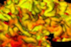
Compared with nonoffenders, violent offenders presented with larger gray-matter volumes bilaterally in the amygdala, the left nucleus accumbens, and the right caudate head. At the same time, the offenders showed smaller gray-matter volumes in the left insula.
Those with substance use disorders demonstrated smaller gray-matter volumes in the orbitofrontal cortex, ventromedial prefrontal cortex, and premotor cortex than men without SUDs.
In addition, regression analysis showed that changes in gray-matter volumes versus the controls corresponded to aggression and psychopathy scores.
"The violent offenders also obtained higher scores than did the nonoffenders for aggressive behavior, and three-quarters of them, as opposed to none of the nonoffenders, met the criteria for [antisocial personality disorder (ASPD)]," Schiffer and colleagues wrote. "The violent offenders obtained higher [Psychopathy Checklist: Screening Version (PCL:SV)] scores than did the nonoffenders, and these scores were comparable to those reported for prison inmates and forensic patients with ASPD."
Finally, the gray-matter volumes in two frontal cortex regions that distinguished those with substance use disorders from nonabusers correlated with response inhibition scores.
"Significant positive correlations between scores for response inhibition and gray-matter volumes of the medial orbitofrontal cortex (r = 0.441, p = 0.002) and the ventromedial prefrontal cortex (r = 0.410, p = 0.004) were observed, indicating that the smaller the volume, the more impaired the ability to withhold responses," Schiffer and colleagues wrote.
The results "confirmed our hypothesis that persistent violent offenders would be characterized by abnormalities of the mesolimbic reward system as evidenced by greater gray-matter volumes in the left nucleus accumbens, the bilateral amygdala, and the right caudate head," they wrote. "Additionally, violent offenders exhibited less gray-matter volume in the left insula."
"Unlike most but not all previous studies, no differences were detected in the gray-matter volume of the prefrontal cortex between the violent offenders and nonoffenders," they wrote.
The results suggest that previous studies of reduced gray-matter volumes in the orbitofrontal cortex, ventromedial prefrontal cortex, and premotor cortex among violent offenders and nonoffenders "may have failed to disentangle the structural brain correlates of persistent violence and SUDs," they wrote, a conclusion supported by two other studies.
Including violent offenders without a history of substance use was a strength of the study but also a limitation, because three-quarters of the "unusual group" met the criteria for antisocial personality disorder, yet had no history of substance use.
"Our study illustrates the use of a strategy for disentangling the correlates of violent behavior from the correlates of SUDs, and if it can be replicated, it may be used in future studies aimed at furthering our understanding of brain mechanisms associated with these two conditions," the authors concluded. The knowledge is needed to help develop effective intervention strategies, as well as hypotheses about the etiology of the disorders, they noted.


