Through the use of 1.5-tesla MRI, Italian researchers have discovered early white-matter brain damage in patients with multiple sclerosis (MS). But the damage did not worsen significantly one year after clinical onset of the disease, according to results published in the November issue of Radiology.
The research, based at the University of Rome "La Sapienza," also showed that gray-matter damage was not detectable at the time of clinical onset, but MRI uncovered a significant decrease in cortical and deep gray-matter volume after one year.
The clinical implications of such brain damage visualized on MRI could play an important role in detecting multiple sclerosis in its early stages. The lead author of the study was Eytan Raz, MD, from the department of neurological sciences (Radiology, November 2010, Vol. 257:2, pp. 448-454).
MRI's capabilities
Although MRI's efficacy in diagnosis, treatment monitoring, and follow-up in patients with multiple sclerosis has been proven in previous research, the connection between clinical signs and radiologic evidence of pathological abnormalities remains unclear. Raz and colleagues noted that this shortfall may be due to gray matter, which is not easily detected by conventional MRI techniques.
"One important question that warrants further study is whether white-matter damage determines abnormalities in gray-matter regions through a disconnection mechanism, or whether gray-matter and white-matter tissues are affected independently," the authors wrote.
To investigate the issue, the researchers consecutively enrolled 34 patients (21 women, 13 men) between 18 and 50 years of age with clinically isolated syndrome (CIS) -- the earliest clinical stage of multiple sclerosis -- at the university's MS center between April 2006 and March 2008. All 34 patients had a single clinical episode indicating multiple sclerosis, including optic neuritis, brainstem syndrome, and spinal cord syndrome.
The study excluded patients who were diagnosed with maladies other than multiple sclerosis, those with presence of other relevant diseases, patients with contraindications to MR imaging, and those who had withdrawn from the study.
All patients underwent a neurologic and an MRI examination at baseline and after 12 months. At the study's one-year follow-up, the mean age of the 34 patients was 32.8 years.
Follow-up scans
Initial and follow-up MRI scans were performed on all patients with a 1.5-tesla system (Gyroscan NT 15, Philips Healthcare, Andover, MA), with a body coil used for signal transmission and a 16-section head coil for sensitivity encoding (SENSE) and for signal reception.
Changes in gray-matter and white-matter integrity were imaged by 3D T1-weighted and diffusion-tensor imaging sequences and by applying voxel-based morphometry (VBM) and tract-based spatial statistics (TBSS) analyses. VBM and TBSS were used to analyze gray-matter volume and white-matter fractional anisotropy, respectively.
At one-year follow-up, MR images revealed that 33 (97%) of the 34 patients exhibited the diagnostic criteria for relapsing multiple sclerosis. Twelve patients (35%) had at least one new relapse during the previous year, while an additional 21 patients (60%), who did not experience a second relapse, showed at least one new T2 lesion in the comparison of follow-up and baseline images. One patient did not satisfy the study's clinical or radiologic criteria for the diagnosis of multiple sclerosis.
Lesion volume
The MR images also revealed that mean T2 lesion volume was 4.37 mL ± 4.2, which was "statistically significantly higher than the baseline values," Raz and colleagues concluded.
The study's longitudinal volumetric analysis showed a significant reduction in global gray-matter volume, while the VBM analysis showed the development of regional gray-matter atrophy in several cortical and subcortical regions in both hemispheres. There was no significant longitudinal change in global or regional white-matter fractional anisotropy.
 |
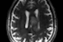
| MR images show regions of decreased gray-matter volume at one-year follow-up in patients with clinically isolated syndrome (CIS). Significant clusters (light blue) of CIS are overlaid on the brain axial sections. Scattered volume reductions are found in several cerebral gray-matter regions, including the thalami, cuneus, paracentral lobule, insula, temporal cortex, caudate head, and supplementary motor areas. Images courtesy of Radiology. |
"It is beyond dispute that the correlation between the one-year follow-up gray-matter volumes and the baseline fractional anisotropy of white matter in our patients is strong," Raz and colleagues wrote. "Gray-matter atrophy increased significantly during the first year after the clinical onset of multiple sclerosis, whereas white-matter damage, which is present from the beginning, did not change significantly in the same time interval."
Varying degrees of combined gray-matter and white-matter damage account for some of the critical changes that occur in the clinical course of patients with multiple sclerosis, the authors wrote.
They recommended that future studies with larger patient populations and extended durations of follow-up should be undertaken for a more definitive conclusion.
By Wayne Forrest
AuntMinnie.com staff writer
November 2, 2010
Related Reading
MRI aids prediction of multiple sclerosis conversion, November 23, 2009
fMRI can help assess therapy for fatigued MS patients, November 10, 2009
Brain iron may play role in multiple sclerosis progression, October 15, 2009
'Benign' multiple sclerosis often evolves to progressive form, February 26, 2007
MRI findings may predict long-term multiple sclerosis disability, January 17, 2002
Copyright © 2010 AuntMinnie.com