The use of intraoperative MRI during pediatric neurosurgery is enabling surgeons at Foothills Medical Centre in Calgary, Alberta, to perform less-invasive procedures by allowing them to modify the surgical plan midsurgery. When using intraoperative MR imaging with integrated neuronavigation capabilities, surgeons can track the extent of the resection of tumors and other lesions.
This ability could potentially increase the safety of tumor resection by eliminating the need for subjective evaluations of anatomical relationships in complex surgical fields and the extent of lesion resection, according to Dr. Mark G. Hamilton, chief of the division of pediatric neurosurgery at Alberta Children's Hospital in Calgary.
Hamilton and colleagues conducted a 10-year retrospective study of a database containing the records of all neurosurgical procedures performed on patients younger than 18 years at Foothills Medical Centre. Their findings, regarding procedures or pathologies in which the use of intraoperative MRI changed the course of surgery or modified surgical strategy, were published in the November issue of the Journal of Neurosurgery: Pediatrics (2009, Vol. 4, pp. 467-474).
The database was initiated in March 1998 and included the records of 98 children, ranging in age from 4 months to 18 years, who had 100 intracranial and five spinal surgical procedures performed through April 2008. The cranial procedures were performed for the treatment of tumors (55%), epilepsy (27%), and vascular lesions (12%). Twenty-two of the procedures were performed for recurrent pathology in 20 patients.
 |
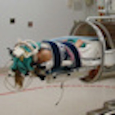
| Photograph of the components of the MR imaging system, including a titanium operating room table, a radiofrequency head coil that can be disassembled, and local radiofrequency shielding. Surgical planning imaging was performed in anesthetized and intubated patients in the operative position. Reprinted from the Journal of Neurosurgery: Pediatrics, with permission from the American Association of Neurological Surgeons. |
At Foothills, the dedicated operating room for MRI-guided surgery contains an actively shielded 1.5-tesla intraoperative MRI mobile scanner (Imris, Winnipeg, Manitoba, Canada) with integrated pointer-based frameless neuronavigation (BrainLab, Feldkirchen, Germany). The magnet moves over the surgical field using overhead crane technology with ceiling-support track beams and an electric motor.
Surgical planning scans were obtained while patients were in the operative position under general anesthesia. These were performed for 97.1% of the procedures in the study, the majority of which (91%) were used for neuronavigation to limit the size of the incision and establish the surgical trajectory.
Intradissection scans were obtained before skin closure, generally before surgeons closed the dura mater, the tough fibrous membrane covering the brain and spinal cord and lining the inner surface of the skull. Images were acquired for 80% of the surgical procedures, adding approximately 30 minutes to the overall operating time.
The authors determined that out of the 84 intradissection scans performed, a significant proportion resulted in further resections to meet the surgeons' operative goals. These ranged from 24% for patients with epilepsy to 49% for patients with tumors. A total of 34 procedures required additional surgery after the images from the intradissection scans were reviewed.
Frequency of intradissection MRI scans by type of tumor or pathology
|
Intradissection scans performed for 46 out of 50 procedures resulted in no further resection but effectively served as quality assurance scans, the authors reported. Twenty quality assurance scans were performed for patients who had additional surgery, and 21 procedures had a postoperative MRI scan only.
The authors emphasized that the study did not evaluate whether the availability of intradissection scanning during surgery improves the extent of tumor resection and/or survival rates in children. Rather, they suggested that the availability of this type of imaging allows a surgeon to temporarily halt surgery to determine if residual disease still exists midway through a complex surgery, and because of this, the surgical procedure could potentially be safer and provide better outcomes for patients.
By Cynthia E. Keen
AuntMinnie.com staff writer
December 8, 2009
Related Reading
Canadian team develops MRI-based robotic neurosurgery device, September 17, 2009
Copyright © 2009 AuntMinnie.com