Researchers at the University of Calgary in Alberta have developed a computerized surgical system called neuroArm that combines MRI and robotics to perform neurosurgery. The system has the potential for expanded microsurgery applications in the future.
The neuroArm allows surgeons to operate in smaller, more intricate areas and potentially could be expanded to other image-guided microsurgical applications. Working on tendons and arteries, other tumors, and surgeries that are not "organ specific" could become "much more straightforward" for surgeons, said Dr. Garnette Sutherland, professor of neurosurgery at the University of Calgary and one of neuroArm's creators.
Early development of neuroArm began about 10 years ago, when the Canadian researchers sought to bring MRI technology to the operating room. "We wanted a high-field MR system that would not impact too much on the practice of neurosurgery nor the nursing, anesthesia, or surgical components," said Sutherland.
They developed an operating suite that featured an MRI scanner, mounted on ceiling rails, that could move in and out of an operating room. The technology was developed further and is now being marketed by intraoperative MRI technology firm IMRIS of Winnipeg, Manitoba. That first prototype system was used in approximately 900 surgical cases, Sutherland estimated.
Design challenges
 |
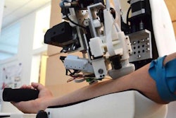
| The robotic neuroArm performs neurosurgery on a patient with a brain tumor. All images courtesy of the University of Calgary. |
Among the design challenges was the basic integration of the MRI technology with the robot. "We didn't want the magnet to affect the robot and we did not want the robot to affect the [MR] images," Sutherland explained. "This robot had to be able to carry an object of about 500 to 750 grams [1.1 to 1.5 lb], had to be able to move that object at 200 millimeters per second, and reach a point within one millimeter."
Researchers chose titanium for much of the system and polyetheretherketone (PEEK) for neuroArm's wrist joints and accessory tools. PEEK is a polymer commonly used in spine fusion applications.
"The reason for [using PEEK] is when you are doing an MR image, you don't get a lot of susceptibility artifact, which you would get with titanium," Sutherland said. "Even though a substance like titanium can go inside an MR machine, titanium can adversely affect the image."
Magnet strength
The prototype magnet strength was 1.5 tesla, but this past summer, the researchers converted to a 3-tesla magnet for integration with the robot. Sutherland said the move to 3 tesla was done to enhance signal-to-noise, "which translates into image quality and speed of image acquisition."
Biopsy or implantation takes place inside the bore of the magnet while MR imaging occurs. Outside the bore, the robot is registered to 3D images acquired intraoperatively to perform the microneurosurgery.
Because a neurosurgeon directs the neuroArm from another room outside of the operating room, the researchers had to "recreate the sound and touch of surgery," Sutherland said. "That was a challenge for our robot. We had to include four sensors that are in contact with the tools that provide a forced feedback to the workstation and the surgeon with the hand controller."
Through that technology, neuroArm can help direct the surgeon on how much pressure to put on a tool, scaling the forced feedback depending on the physical characteristics of what the tool touches.
 |
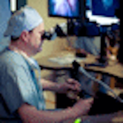
| The neuroArm's intuitive sensors help Dr. Garnette Sutherland get a feel for the surgical procedure as he operates from another room. |
So far, neuroArm has been in six surgical cases for brain tumors of various types, with Sutherland as the sole practitioner. The university currently is developing a training program for other surgeons. "I would say [neuroArm] is relatively intuitive and the surgeon should be able to cover most of the knowledge base and practice on the robot in about two weeks," Sutherland added.
Meanwhile, the Canadian researchers are moving toward their goal of a 100-patient clinical study to demonstrate neuroArm's value.
"As genetic people become wiser and give us tools to change cells, surgery will shift to a much more minimalistic approach, which would include implantation into body parts," Sutherland said. "What better way to do that than with a robot that is image-guided?"
By Wayne Forrest
AuntMinnie.com staff writer
September 17, 2009
Related Reading
IMRIS gets FDA nod, September 8, 2009
Swiss group performs transcranial HIFU, June 23, 2009
IMRIS wins CE Mark for MR angiography suite, May 1, 2009
Gestix device gives brain surgeons a hand in the OR, July 31, 2008
Copyright © 2009 AuntMinnie.com