Dynamic contrast MRI has been used to try to characterize breast tumors, and it has been studied for links between its findings and prognostic indicators in breast cancer. But there has not been sufficient clinical research that explores the prognostic value of breast MR imaging for classifying patients into good versus poor treatment outcomes, according to researchers at the University of California, San Francisco and the University of Washington in Seattle.
High-spatial-resolution signal enhancement ratio (SER) imaging can show the heterogeneous microvascular network in breast cancers, wrote Ka-Loh Li, Ph.D., and colleagues in a study published in Radiology (July 2008, Vol. 248:1, pp. 79-87). Li's team assessed this type of imaging's ability to predict disease recurrence in patients with breast cancer who had preoperative MR exams.
The group used gadolinium-enhanced MR imaging data taken for 48 women with invasive breast cancer who preoperatively had had four cycles of therapy between 1995 and 2002. Contrast MR imaging was performed before and after the chemotherapy on a 1.5-tesla device (Signa, GE Healthcare, Chalfont St. Giles, U.K.); three phased-array breast coils, a closed breast coil (also GE), and two open breast coils (MRI Devices, Waukesha, WI) were used. The data were taken at three time points: one precontrast acquisition followed by two postcontrast acquisitions in two consecutive five-minute intervals.
The women were divided into two groups: recurrence and recurrence-free. Li's team defined cutoff criteria to predict disease recurrence after surgery and established the following tumor volume set ranges of SER values from high-spatial-resolution dynamic contrast-enhanced MR imaging data:
- 0.47 to 0.71 (low SER)
- Greater than 0.71 to 1.45
- Greater than 1.45 (high SER)
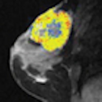
SER maps from a patient in the recurrence-free group showed that chemotherapy reduced tumor volume, especially the number of voxels with high SER values:
 |
| MR imaging study results before and after four cycles of chemotherapy in a 39-year-old woman in the recurrence-free group. Representative color-coded SER maps overlaid on corresponding grayscale images of first postcontrast acquisition (three-dimensional fast gradient-recalled echo, 8/4.2, 20° flip angle) before (top row) and after (bottom row) chemotherapy. Fig. 1a. Li KL, Partridge SC, Joe BN, et al. "Invasive Breast Cancer: Predicting Disease Recurrence by Using High-Spatial-Resolution Signal Enhancement Ratio Imaging." Radiology. 2008;248(1):79-87. All images courtesy of RSNA. |
Three prechemotherapy parameters -- total tumor volume and tumor volumes with high and low SER -- and the postchemotherapy tumor volume with high SER differed between patients with recurrence and those without.
 |
| Comparison of the number of voxels with high (hypothesis 1) and low (hypothesis 2) SER between recurrence-free group (group 1) and recurrence group (group 2). Box-and-whisker plots (a-c) before chemotherapy and (d-f) after chemotherapy. (a, d) SER is between 0.47 and 0.71. (b, e) SER is greater than 1.45. (c, f) All voxels with nonzero SER. Interquartile range (i.e., 25%-75%) is shown by box, extreme values are shown by whiskers, and median is shown by bar within the box. P-value of the difference between the two groups was evaluated with Wilcoxon rank sum test. Dashed lines = cutoff criteria. Fig. 3a-f. Li KL, Partridge SC, Joe BN, et al. "Invasive Breast Cancer: Predicting Disease Recurrence by Using High-Spatial-Resolution Signal Enhancement Ratio Imaging." Radiology. 2008;248(1):79-87. |
The researchers found that the size of the segmented tumor volume with high SER could predict tumor recurrence, and that the size of tumor volume in voxels of cancerous tissues infiltrating into the breast parenchyma could also predict cancer recurrence. They concluded that combining the use of SER predictors with traditional prognostic factors could improve the prediction of prognosis for patients at high risk of disease recurrence and death.
By Kate Madden Yee
AuntMinnie.com staff writer
July 2, 2008
Related Reading
Contrast MRI helps track breast cancer therapy response, June 6, 2008
Breast MRI with USPIO contrast helps assess lymph node metastases, April 25, 2008
Reimbursement crunch throttles breast MRI CAD users, April 8, 2008
MRI beats mammography, US in detecting breast cancer in high-risk women, March 12, 2008
Catching undetected cancer with MR plus mammo in high-risk women outweighs costs, December 17, 2007
Copyright © 2008 AuntMinnie.com