Dear MRI Insider,
One of the few truly entertaining shows on the misnamed Music Television (MTV) network is "Pimp My Ride," in which ordinary folks have their equally average cars souped up, tricked out, and customized until they are the proud owners of the biggest, baddest vehicles on the block.
In a way, MR has undergone a similar flashy transformation with amped up magnets and showy imaging sequences. In fact, we look forward to the day when a vendor hosts a "Pimp My MRI" promotion at RSNA.
Until then, we offer a number of articles on some of the latest advances in MRI. First, our Insider Exclusive is a contribution from E. Mark Haacke, Ph.D., director of the MRI Institute for Biomedical Research at Wayne State University in Detroit. He is revved up about susceptibility-weighted imaging (SWI) and its myriad applications in neuroimaging. Click here to read more.
We also have the results of an in vitro study that evaluated the relaxivity rates associated with different gadolinium-based contrast agents. The researchers conducted their experiment on 1.5- and 3-tesla scanners.
Speaking of fast and furious magnets, the gentlemen at MRIPlanning.com have something to say about the tesla wars. If a facility opts for a higher field strength scanner, what does that mean from a siting point of view? Learn the answer by cruising back to the MRI Digital Community on Tuesday.
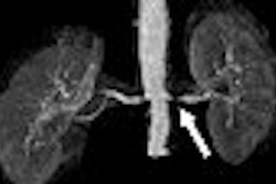
You can also learn about how invaluable interoperative MR is for neurosurgical and ENT procedures, how MR can assess whether arterial stiffness is tied to mortality in pulmonary hypertension, and why brain microbleeds on MR are associated with higher ambulatory blood pressure. Finally, results from a functional MRI study may hold a key as to why people are driven to overeat.