The latest news in breast MR: Negative MR findings should not be considered a marker for benign lesions in microcalcifications initially found on mammography. On the other hand, adding computer-aided detection (CAD) to breast MRI can boost specificity without decreasing its sensitivity for cancer detection.
In the first study, researchers with the Italian Trial for Breast MRI of Microcalcifications tested dynamic MRI for evaluating microcalcifications that were deemed suspicious on mammography. These microcalcifications are a common feature of early breast lesions and are sometimes the only sign in clinically occult cancer, explained Dr. Massimo Bazzocchi and colleagues in the American Journal of Roentgenology.
Bazzocchi is from the University of Udine in Udine, Italy. His co-authors are from multiple Italian institutions, including the Istituto Policlinico San Donato in Milan and the University of Tor Vergata in Rome.
"The role of MRI in characterizing breast microcalcifications remains a debated issue. The reported values of sensitivity range between 45% and 100%, and the specificity between 37% and 95%," the authors wrote. "Our results showed a relatively low sensitivity (87%) of breast MRI for the detection of breast cancers associated with microcalcifications" (AJR, June 2006, Vol. 186:6, pp. 1723-1732).
For this study, 17 centers enrolled 174 patients from 1998 to 2000. Imaging was done on a 1- or 1.5-tesla MRI unit 30 days after standard mammography. Dynamic MRI was performed on the seventh to 14th day of the menstrual cycle with a 0.1 mmol/kg injection of gadoteridol (ProHance, Bracco, Milan, Italy). Surgical biopsy was done two weeks after MR. Lesions were classified as benign or malignant.
 |
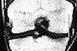
| Ductal carcinoma in situ in left breast of 44-year-old woman. Oblique view mammogram and mammographic magnification view of surgical specimen shows cluster of punctate calcifications (arrow) close to hookwire. |
 |
Of the 112 lesions, 33 were ductal carcinoma in situ (DCIS) and 42 were either invasive ductal carcinoma (IDC) or invasive lobular carcinoma (ILC). All presented as microcalcification clusters with and without opacity.
The diagnosis of DCIS was confirmed by pathology, and 87.8% of those 33 lesions showed enhancement after contrast agent injection, the authors stated in their results. There were seven false-negative cases, all of which had presented mammographically as microcalcifications. The sensitivity of MRI for DCIS was 79%.
For the 42 invasive carcinomas, 95.2% showed enhancement on MR. The two false-negative cases were a mixed ILC-lobular carcinoma in situ and one mixed DCIS-IDC. Both also presented as a cluster of microcalcifications on mammography. The sensitivity of MRI for invasive cancer was 93%.
 |
| Same patient as above. The MR study (axial plane) does not show any foci of enhancement. Bazzocchi M, Zuiani C, Panizza P, Del Frate C, Soldano F, Isola M, Sardanelli F, Giuseppetti GM, Simonetti G, Lattanzio V, Del Maschio A, "Contrast-Enhanced Breast MRI in Patients with Suspicious Microcalcifications on Mammography: Results of a Multicenter Trial" (AJR 2006; 186:1723-1732). |
Overall, the sensitivity of MRI was 87%, the specificity was 68%, the positive predictive value was 84%, the negative predictive value was 71%, and the accuracy was 80%.
"The not-perfect sensitivity of MRI -- 87% -- is a crucial point that prevents us from clinical use of MRI in the diagnosis of mammographically detected microcalcifications," the group wrote. "MRI cannot replace percutaneous or surgical biopsy, which gives pathological information."
However, the authors added that MRI did demonstrate a relatively good specificity at 68% and an overall accuracy of 80%. They credited technological improvements, such as use of a 3D sequence, for this uptick in performance.
Breast MRI with CAD
In another new study, Dr. Constance Lehman and colleagues from the University of Washington in Seattle and the Seattle Cancer Care Alliance compared the accuracy of breast MRI interpretation with and without a CAD system (CADstream, Confirma, Kirkland, WA).
"Our findings suggest this software may improve the accuracy of radiologists' interpretations of enhancing lesions identified as suspicious on breast MRI," wrote Lehman's group (AJR, July 2006, No. 187:1, pp. 51-56).
The study included all lesions biopsied with MR-guidance between March 2001 and August 2002, resulting in 33 lesions in 29 female patients. MR was done on a 1.5-tesla scanner with a dedicated breast coil (LX, GE Healthcare, Chalfont St. Giles, U.K; 1.5T 4 Channel Breast Array, MRI Devices, Waukesha, WI). All MR exams were retrospectively processed by the CAD system, which incorporate three MR series into its calculations:
- An unenhanced T1-weighted series
- An immediate contrast-enhanced T1-weighted series
- A delayed contrast-enhanced T1-weighted series
One radiologist recorded whether the study lesion was marked as having significant enhancement by CAD at 50%, 80%, and 100% enhancement threshold levels. For each lesion significant enhancement, the percent values of washout, plateau, and persistent enhancement were also recorded.
According to the results, 24 lesions were identified by pathology as benign and nine were malignant. "All malignant lesions showed significant enhancement using the program at all thresholds, producing a sensitivity of 100%," the authors wrote.
"False-positive rates for the computer-aided assessment compared with the original radiologist assessment were reduced by 25% at a 50% threshold, 33% at an 80% threshold, and 50% at a 100% threshold for enhancement," they added.
 |
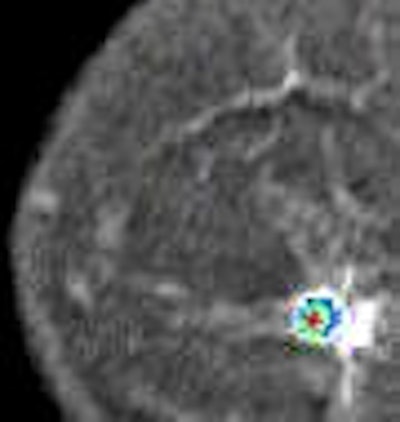
| A 49-year-old woman with mammographically occult contralateral breast cancer detected on MRI. Above, initial contrast-enhanced sagittal MR image. Below, CAD overlay shows lesion as having significant enhancement and mixed pattern of washout, plateau, and persistent delayed enhancement. If pixel value on delayed series decreases by more than 10% of its immediate contrast-enhanced value, that pixel is color-coded red on monitor, indicating washout of contrast material. If pixel value increases by more than 10%, it is color-coded blue on monitor, indicating persistent enhancement. If pixel value does not change in either direction by more than 10%, it is color-coded green for plateau enhancement. |
 |
| Lehman CD, Peacock S, DeMartini WB, Chen X, "A New Automated Software System to Evaluate Breast MR Examinations: Improved Specificity Without Decreased Sensitivity" (AJR 2006; 187:51-56). |
In terms of kinetic enhancement pattern types in eight benign and eight malignant lesions, all showed a wide range of washout, persistent, and plateau. There was a significant overlap of benign and malignant lesions.
Lehman's group stated that they found the information from the initial portion of the time-signal intensity curve provided by CAD to be the best predictor of malignancy. Their results do differ from a previous study by Dr. Christiane Kuhl and colleagues that reported delayed contrast activity was predictive of histology (Radiology, April 1999, Vol. 211:1, pp. 101-110).
The authors of the current study offered some reasons for these differing results: the CAD system used two contrast-enhanced series and provided detailed information about each pixel in a given tumor. Earlier technology required that the reader place a region of interest on the most suspicious area after visual inspection.
They acknowledged that this study was done with certain limitations including the single-site, retrospective design. Also, there is currently no clear consensus among breast imaging experts as how best to acquire MR images, which could affect how the CAD performs, they added.
However, the group deemed these preliminary results and promising. "If these results are validated by a larger study, the number of unnecessary biopsies of MR lesions could be reduced without a concomitant decrease in cancer detection, they stated. "These (CAD) programs may decrease heterogeneity of interpretations across radiologists of varying levels of experience in breast MR interpretations."
By Shalmali Pal
AuntMinnie.com staff writer
July 3, 2006
Related Reading
MR spectroscopy aids breast cancer diagnosis, June 8, 2006
Technology, guidelines cement role for breast MR in cancer screening, May 26, 2006
CAD finds breast cancers that radiologists missed for years, March 6, 2006
MR mammography: Screening tool or diagnostic adjunct? February 9, 2006
Larger lesions on breast MRI equal greater chance of cancer, February 8, 2006
Copyright © 2006 AuntMinnie.com