Mental health specialists once relied heavily on postmortem studies to document abnormal brain structures in schizophrenia. This early research led to the hypotheses that schizophrenics had no structural brain abnormalities when compared to normal subjects.
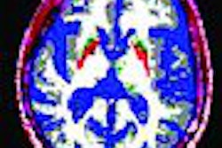
This incorrect notion prevailed as late as the 20th century until the advent of CT and MRI revealed a clearer picture of the schizophrenic brain. Early CT studies indicated large lateral ventricles in schizophrenia. MRI studies revealed glaring distinctions between gray and white matter (Surgical Planning Laboratory, Harvard University, February 3, 2004).
Fast-forward to 2006 with two new MRI studies adding to the literature on brain abnormalities in schizophrenia. The first, out of Australia, took a closer look at brain volume before and after the onset of illness. The second paper by U.S. researchers mapped gray-matter loss rates in childhood-onset schizophrenia.
Volume, stage, diagnosis
Dr. Dennis Velakoulis and colleagues addressed the specificity and timing of hippocampal and amygdala volume changes in three groups of patients: those with first-episode psychoses, those with chronic schizophrenia, and those identified as having an ultrahigh risk (UHR) for psychosis. There was also a fourth control group.
Velakoulis and several co-investigators are from the University of Melbourne. Other contributors are from the Royal Melbourne Hospital, the Brain Research Institute, and the Howard Florey Institute, all in Melbourne.
The study population consisted of 473 individuals: 89 with chronic schizophrenia, 162 with first-episode psychosis, 135 with a UHR for psychosis, and 87 controls. The first-episode patients were subdivided into those with first-episode schizophreniform psychosis, those with affective psychosis, and those with other psychoses. Of the 135 UHR patients, 39 subsequently developed a psychotic illness (UHR-P) and 96 did not (UHR-NP).
All imaging was done on a 1.5-tesla scanner (Signa, GE Healthcare, Chalfont St. Giles, U.K.) using a 3D volumetric spoiled gradient-recalled echo, steady-state sequence. Hippocampal volumes were estimates using a manual tracing technique and predefined anatomic criteria. Amygdala volumes were assessed using a method that separated it from the adjoining hippocampal formation. A 3D morphometric approach was taken for whole-brain volumes, and the intracranial volumes (ICV) were estimated from a sagittal reformat of the original 3D MR dataset.
The results showed that hippocampal volumes were reduced bilaterally in patients with chronic schizophrenia and on the left side in first-episode patients. First-episode schizophreniform patients showed no volume reduction, and neither the UHR-P nor UHR-NP groups saw a reduction in hippocampal volumes.
Meanwhile, in the amygdala, volumes were larger only in patients with first-episode nonschizophrenic psychoses. Amygdala volumes did not differ among the other groups when compared to the controls.
Finally, whole-brain volumes were smaller in patients with first-episode schizophreniform disorder, as well as in patients with first-episode affective psychoses and the UHR-NP group. There were no significant ICV differences among all the groups.
The authors summarized that "patients with chronic schizophrenia have a bilaterally smaller hippocampi; that left-sided hippocampal volume reduction is seen in first-episode patients with schizophrenia but not in those with schizophreniform, affective psychoses, or other psychoses; and that bilateral amygdala enlargement was present only in the first-episode nonschizophrenia patients" (Archives of General Psychiatry, February 2006, Vol. 63:2, pp. 139-149).
A lack of change in hippocampal volumes in first-episode schizophreniform psychosis is especially important because this group differed from patients with first-episode schizophrenia in illness duration but not in other areas such as age, gender, or time of presentation to MRI, they noted.
These findings support earlier studies that indicate hippocampal volume reduction is a marker of illness rather than a marker of risk for illness, and that altered volumes are not a premorbid marker of vulnerability for psychosis, they stated.
"Our data provide further evidence that structural medial temporal changes occur during and immediately after the transition to first-episode psychosis and provide an impetus for further studies in the neurobiologic process of early psychosis," they wrote.
Gray-matter loss
In a second study, a group from the University of California, Los Angeles (UCLA), and the National Institutes of Health (NIH) in Bethesda, MD, mapped out the pattern of gray-matter loss in childhood-onset schizophrenia (COS), in which psychotic symptoms manifest by age 12.
For this five-year longitudinal study, lead author Christine Vidal, Ph.D., and colleagues looked specifically at the effect of the gray-matter deficit trajectory on the medial hemispheric surface and key frontal and limbic regions implicated in schizophrenia.
The study involved 33 subjects: 12 children with schizophrenia, 12 healthy controls, and nine kids with psychosis not otherwise specified (PNOS) but who were on medication. The latter were included to assess if neuroleptics played a role in progressive gray-matter deficit, the authors explained.
All subjects underwent 3D MRI on a 1.5-tesla scanner (Signa, GE Healthcare). The protocol included T1-weighted fast spoiled gradient-echo imaging. Several different kinds of maps were produced using the MR information: tissue maps, 3D cortical maps, cortical pattern matching to localize disease effects on cortical anatomy over time, matching cortical anatomy maps to match gyral patterns across all subjects, and gray-matter loss maps.
"Three-dimensional maps of brain changes, derived from the same subjects over time, showed profound, progressive gray-matter loss in some medial walls regions in schizophrenia," the authors stated of their findings. "In schizophrenic subjects, a striking gray-matter loss was observed in superior medial frontal regions including the superior frontal and precentral gyri" (Archives of General Psychiatry, January 2006, Vol. 63:1, pp. 25-34).
Maps of gray-matter loss confirmed those findings, again showing highly significant, progressive deficit localized in the superior medial front cortex of the COS subjects, but little loss in the normal adolescents.
To detect how early these losses occurred, the authors compared gray-matter profiles across 24 subjects at their first scan and their last, 4.6 years later. They found that the cingulate was relatively preserved at the onset of disease but showed significant deficits in the left hemisphere at follow-up.
For the PNOS group, the authors determined that these subjects who did not meet the clinical criteria for schizophrenia still shared some of the same deficits of the condition, including significantly accelerated gray-matter loss in the superior medial frontal and cingulate areas. However, their symptoms were less pervasive than in the COS children. These findings support the theory that the progressive changes in COS are not attributable to purely psychiatric illness.
"The longitudinal brain maps show (that) a sharp boundary in the profile of gray-matter loss is visible between frontal and limbic tissue, suggesting differential susceptibility," they wrote. "The overall profile of progressive frontal gray-matter loss reinforces the notion that COS is neurobiologically continuous with the later-onset illness."
These dynamic maps of schizophrenia, demonstrating that frontal and limbic regions may not be equally vulnerable to gray-matter attrition, could serve as a predictive tool to determine if someone is seriously at risk for the disease, the authors added.
By Shalmali Pal
AuntMinnie.com staff writer
April 4, 2006
Related Reading
DTI scans track schizophrenia-like changes in brains of teen marijuana users, December 1, 2005
MRI studies make sense of multifaceted schizophrenia, April 27, 2005
Olanzapine has various effects on striatal volumes in schizophrenia, October 26, 2004
Copyright © 2006 AuntMinnie.com


