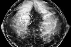
Pregnancy-associated breast cancer is often found at an advanced stage, partly because of decreased mammographic sensitivity in the lactating breast. A report released today in Radiology describes how MRI can cut through the increased parenchymal density, glandular volume, and water content of the lactating breast.
Leandro Espinosa and colleagues at Stanford University in Stanford, CA, retrospectively reviewed cases at their institution and compiled a list of MRI characteristics of breast cancer in lactating women. Espinosa's co-authors included Drs. Bruce Daniel and Debra Ikeda.
Seven women were included in their final review, all of whom were breast-feeding at the time of the MR exam or had been breast-feeding a month before. While lactating, they underwent unilateral or bilateral breast MRI for biopsy-proven palpable cancer, angiosarcoma, or a strong family history.
Imaging was done on a 1.5-tesla MR scanner with a dedicated breast coil (EchoSpeed, GE Healthcare, Chalfont St. Giles, UK; MR Devices, Waukesha, WI). The protocol included T1-weighted spin-echo imaging, nonenhanced T2-weighted 3D imaging, and dynamic T1-weighted imaging with gadolinium.
Readers came to a consensus when analyzing the images, looking at the signal intensity and overall appearance of glandular tissue, potential patterns of enhancement, and morphologic features.
The results showed that all seven patients had multiple dilated ducts with high T2-weighted imaging signal intensity. This was consistent with the presence of milk. Imaging of all patients also showed rapid initial contrast enhancement, a type 3 or type 5 average signal intensity curve, and a one-minute change in signal intensity of 38%. Six of the women had uniformly high glandular tissue signal intensity on T2-weighted images.
Ultimately, five out of the seven women were diagnosed with ductal carcinoma in situ (DCIS). "At nonenhanced T2-weighted MR imaging, the tumors in all five patients had low signal intensity compared with the normal lactating glandular tissue," the authors wrote. "At contrast-enhanced T1-weighted MR imaging, all invasive tumors in all five patients were visible as focal-enhancing lesions" (Radiology, November 2005, Vol. 237:2, pp. 429-436).
The authors concluded that the following were MR characteristics of normal lactating breast tissue:
- Increased glandular density
- High T2-weighted signal intensity
- Rapid but moderate contrast enhancement
In comparison, MR findings of DCIS in lactating women included the following:
- Lower T2-weighted signal intensity throughout the DCIS
- High signal intensity in the uninvolved lactating tissue
- More intense initial contrast enhancement
- Early washout
The researchers noted that all the tumors in their series were visible to some degree on mammography or ultrasound. However, they stated that MR may be more reliable for depicting the extent of invasive carcinomas in lactating patients.
Imaging safety in pregnant and breast-feeding women will be the topic of discussion at a refresher course (RC807) at the 2005 RSNA in Chicago. "Issues and Controversies in Imaging the Pregnant Patient" will address the use of iodinated and gadolinium contrast media in these patients. A separate scientific presentation (SSJ02-05) will showcase the range of sonographic signs and symptoms found in lactating women with a suspected breast abscess.
By Shalmali Pal
AuntMinnie.com staff writer
October 24, 2005
Related Reading
Breast MR helps refine partial-breast irradiation, October 19, 2005
MR spectroscopy aids breast MR imaging, July 29, 2005
Breast abscesses in the clear with ultrasound-guided drainage, September 2, 2004
Copyright © 2005 AuntMinnie.com