When combined with MR, diffusion-weighted imaging (DWI) produces a quantitative biophysical parameter called the apparent diffusion coefficient (ADC) of water. This ADC is indicative of the movement of water within the tissue.
As cells attempt to maintain homeostasis, water flows in, out, and around the cells, thereby increasing or decreasing the ADC value. ADC maps have been successfully employed to monitor the progression of ischemic tissue in stroke. Imaging and interventional specialists are now proposing the use of ADC maps to monitor uterine fibroid therapy.
Diffusion-weighted MRI for US therapy
"The findings of this study support the hypothesis that a multiparametric approach by using DW imaging, ADC mapping, and conventional MR imaging can accurately be used to identify ablated uterine tissue after focus ultrasound surgical treatment," wrote lead author Michael Jacobs, Ph.D., from the Johns Hopkins University School of Medicine in Baltimore (Radiology, July 2005, Vol. 236:1, pp. 196-203).
Co-author Dr. Hyung Kim is from the Johns Hopkins University School of Medicine in Baltimore; co-author Dr. Edward Herskovits, Ph.D., is from the University of Pennsylvania in Philadelphia.
For their prospective feasibility study, 14 patients with symptomatic fibroids were enrolled. Fibroids deemed responsible for clinical symptoms were identified with pretreatment T2-weighted and T1-weighted contrast-enhanced MR. Treatment was done with an ultrasound surgical system (TX Sonics, InSightec, Haifa, Israel) and a 1.5-tesla MRI (Excite, GE Healthcare, Chalfont St. Giles, U.K.).
The patients underwent MR exams in a prone position. The imaging sequence included phase-sensitive T1-weighted fast spoiled gradient-recalled MR. Images were acquired after each round of MR image-guided focused ultrasound therapy. Post-treatment verification of ablated tissue consisted of T1-weigthed fast spoiled gradient MR with and without contrast. For DWI, regions of interest were drawn to include 50% to 80% of the treated fibroid.
Before ultrasound treatment, contrast-enhancing fibroids had no discernible signal intensity changes on conventional MR, DWI, or ADC maps. However, after treatment, DWI in all patients demonstrated increased signal intensity changes localized in treated fibroid regions, the authors reported.
 |
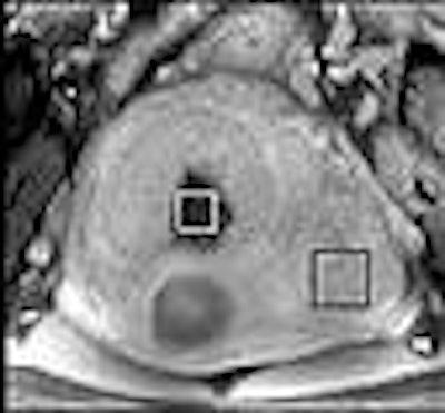
| Representative coronal postcontrast T1-weighted MR images (Contrast T1), DW images (DWI), T2-weighted MR images (T2WI), and ADC maps acquired in 48-year-old woman. Arrows show location of fibroid, and squares demonstrate the region of interest selected for quantification of ADC values. Row A, Enhancing fibroids were seen on baseline T1-weighted MR image (185/1.5) with no discernible signal changes on DW image (5000/90, b = 1000) or T2-weighted MR image (5000/90, b = 0). ADC map shows no discernible signal changes. Row B, after treatment, postcontrast T1-weighted MR image demonstrates area of hypointensity within the treated region, with increased signal intensity in the same regions on the DW image and corresponding decreased signal intensity on ADC map. Row C, at six-month follow-up, a persistent area of hypointensity is noted in the area of treated fibroid as shown on the T1-weighted postcontrast image. The DW image signal intensity is heterogeneous with the treated region, and the central region shows increased signal intensity on the ADC map that is colocalized with the T1-weighted MR image. Figure 2. Jacobs MA, Herskovits EH, Kim HS. Uterine fibroids: diffusion-weighted MR imaging for monitoring therapy with focused ultrasound surgery -- preliminary study. Radiology 2005;236:196-203. |
"The signal intensity on DW images appears heterogeneous within the treated region, and increased signal intensity is exhibited on the ADC map," they wrote, adding that their study did "demonstrate the ability of DW imaging to ADC mapping to demarcate regional diffusion abnormalities over a temporal period."
The mean ADC value at baseline in the fibroids was 1553 mm-6/sec2. The mean ADC value was significant reduced in treated fibroids (1078 mm-6/sec2) in comparison to untreated ones (1685 mm-6/sec2). Finally, there was a significant difference between the mean post-treated ADC value (1905 mm-6/sec2) and the ADC value of untreated fibroids (1437 mm-6/sec2).
"It is believed that the ADC can be used to measure cytotoxic edema caused by swelling of cells, tortuosity of the path of diffusion, and structural dehydrating of the fibers, and is very sensitive to the acute changes in water in tissue," the authors stated.
The biggest advantage of DWI is that it is a noninvasive exam, decreasing the chance of a negative reaction to contrast agent. In addition, DWI is done with the patient under conscious sedation, which can cut back on artifacts.
Diffusion-weighted MRI for UFE
In a previous paper, Jacobs, Kim, and others from Johns Hopkins also found positive results with diffusion-weighted echoplanar MRI for assessing fibroid and myometrium response to uterine fibroid embolization (UFE) (Journal of Computer Assisted Tomography, January/February 2005, Vol. 28:1, pp. 83-86).
Their hypothesis in this study was that reducing or eliminating blood flow to the treated fibroids would lead to membranous destruction and cellular dehydration, indicating that the UFE had been successful, explained lead author Dr. Eleni Liapi.
For this retrospective study, the group found 11 premenopausal women (32 fibroids) who underwent UFE and diffusion-weighted MRI between April 2002 and March 2003. They were scanned on a 1.5-tesla unit (CV/i, GE Healthcare). The protocol included T2-weighted FSE images and breath-hold images without contrast and with 0.1 mmol/kg of gadodiamide (Omniscan, GE Biosciences).
The DWI series was done with breath-hold and was performed in the axial plane along with echoplanar imaging. Fat suppression was added to avoid chemical shift artifacts. Two radiologists in consensus performed image analysis. The imaging variables that were assessed before and after UFE included maximum diameter, signal intensity, and degree of enhancement.
ADC maps were used for quantitative measurements. "The ADC maps that are generated using DWI provide accurate quantification of the cellular motion of water molecules," the authors explained.
On the pretreatment T1 sequence, 97% of the lesions were isointense in comparison to muscle. The mean lesion diameter pre-UFE was 5 cm, which decreased by 1 cm after embolization. The mean fibroid enhancement was 82% before UFE and 11% after UFE.
Before treatment, the mean ADC value of fibroids was 1.74 E-3 mm2/s. The ADC decreased to 1.22 E-3 mm2/s afterward. The mean ADC value of normal myometrial tissue before UFE was 2.05 E-3 mm2/s and 1.96 E-3 mm2/s afterward.
"According to these preliminary results, DWI and ADC maps may depict specific changes in water composition of uterine fibroids after embolization," the authors concluded. "Treated fibroids had low ADC values after treatment, compared with ADC values before treatment. ADC values for the myometrium remained stable, indicating specific treatment response."
By Shalmali Pal
AuntMinnie.com staff writer
September 9, 2005
Related Reading
ACOG study shows UFE is more cost-effective than surgery, June 17, 2005
Post-UFE complications rare, but may require more serious surgery, June 13, 2005
Susceptibility-weighted MRI ups contrast, offers minute detail, September 15, 2004
Copyright © 2005 AuntMinnie.com