Fast spin-echo MRI turned in a lower sensitivity than conventional spin-echo for evaluating meniscal tears, according to a study out of Duke University in Durham, NC. As a result, the researchers recommended that musculoskeletal radiologists "abandon" fast-spin echo MRI in the meniscus.
The sensitivity of fast-spin echo MRI was 80% compared to 90% for conventional spin-echo, according to the study in the American Journal of Roentgenology by Drs. Garyun Blackmon, Nancy Major, and Clyde Helms. They used both conventional spin-echo and fast-spin echo sequences to detect tears in 432 menisci in 216 patients. The authors noted that they would have failed to detect 42 tears among the 432 menisci if they had relied on fast-spin echo sequences alone.
Imaging parameters for conventional spin-echo MRI were as follows:
- TR/TE 2000/20
- Matrix 256 x 192 signal averages
- 16-cm field-of-view
- 4-mm slices with a 0.4-mm gap
- Imaging time of seven minutes and 20 seconds
For the fast-spin echo sequences, imaging parameters were the same except for TR/TEeff of 3,000/17 and echo-train length of 4. Imaging time was three minutes and 20 seconds.
Sagittal images were used for evaluating the menisci, and both sequences were proton density-weighted with fat suppression. Imaging was done on a 1.5-tesla system (Signa, GE Healthcare, Chalfont St. Giles, UK).
 |
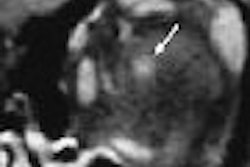
| Conventional spin-echo versus fast spin-echo imaging for detection of meniscal tear in 50-year-old woman. Sagittal conventional spin-echo proton density-weighted MR image (TR/TE, 2,000/20) with fat suppression obtained through medial meniscus shows complex tear of posterior horn (arrow). |
Conventional spin-echo detected 170 tears, while fast-spin echo detected 128. The findings were also discordant for 72 menisci, the researchers reported. The research group reviewed five other published studies and noted, "the varying opinions regarding the utility of fast spin-echo sequences seem to be based on different conclusions derived from the studies rather than differences in the results generated from the studies, because all show an approximately 80% sensitivity for meniscal tears with the fast spin-echo sequence" (AJR, June 2005, Vol. 184, pp. 1740-1743).
The sensitivity of conventional spin-echo imaging was determined from a control of group of 64 patients in whom 128 menisci were examined for tears using arthroscopy as the gold standard. In this group, 49 of 53 tears were detected by conventional spin-echo imaging, resulting in a sensitivity of 93%. There were two false-positive cases, resulting in a specificity of 97%.
 |
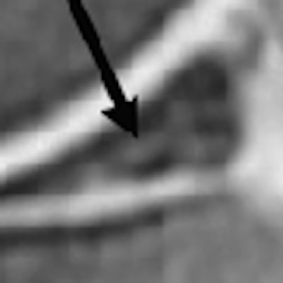
| Conventional spin-echo versus fast spin-echo imaging for detection of meniscal tear in 50-year-old woman. Sagittal fast spin-echo proton density-weighted MR image (3,000/17) with fat suppression obtained through medial meniscus does not show meniscal tear. Garyun BB, Major NM, Helms CA, "Comparison of Fast Spin-Echo Versus Conventional Spin-Echo MRI for Evaluating Meniscal Tears" (AJR 2005; 184:1740-1743). |
"Taking into account the expected false negatives and false positives generated from the control group, we found that the sensitivity of the fast spin-echo sequence was 80%," the authors explained.
In the 17% of cases in which the findings from conventional spin-echo and fast spin-echo imaging were discordant, consensus opinion was reached after re-examination by two of the participating radiologists.
Forty-two of the discordant interpretations were because of tears that were readily detected on conventional spin-echo, but not identified on fast spin-echo imaging. Twenty-seven of these were medial tears and 15 lateral tears.
In the remaining 30 discordant interpretations, abnormal meniscal signals present on fast spin-echo images were not present on conventional spin-echo images.
"Both the group of false-positive and false-negative meniscal tears on fast spin-echo imaging may be attributed in part to blurring," the researchers wrote, explaining that blurring was more evident with the short-TEeff sequences.
"We strongly urge abandonment of fast spin-echo sequences for evaluating the menisci," the authors concluded. However, a prospective study directly comparing conventional spin-echo and fast spin-echo sequences with arthroscopy would provide more precise data, they wrote.
By N. Shivapriya
AuntMinnie.com contributing writer
July 6 2005
Related Reading
Meniscal MRI studies highlight axial advantages, radial tear rates, August 28, 2004
MRI pinpoints meniscal tears, proves less valuable for knee trauma, January 9, 2003
Copyright © 2005 AuntMinnie.com