In traditional MRI, both cerebral abscesses and high-grade tumors can be depicted as rim-enhancing lesions, rendering them indistinguishable from each other. PET techniques can help tell them apart, often showing increased metabolism in high-grade tumors and decreased metabolism in abscesses.
But even when PET is available, it makes sense for patients undergoing MR evaluation of a brain mass to use a second MR technique if the first is nondiagnostic. Some have advocated the use of MR spectroscopy for this purpose, but its use with abscesses has been limited.
Researchers at Duke University in Durham, NC, had another idea. They posited that because high-grade tumors often show increased vascularity relative to normal parenchyma, measuring relative blood volumes of the two lesion types with MR perfusion imaging might unveil their true identities.
At the 2000 RSNA meeting, Duke University Medical Center associate professor of radiology Dr. James Provenzale described a study that found distinct hemodynamic differences between the two lesion types.
"The purpose of this study was to determine if hemodynamic measurements using MR blood volume imaging could be used to distinguish rim-enhancing neoplasms from cerebral abscesses," Provenzale said.
The study looked at 7 patients, 4 of whom had biopsy-proven rim-enhancing high-grade gliomas (average age 47). This group included two patients with glioblastoma multiforme, one with gliosarcoma, and one with anaplastic astrocytoma.
The remaining 3 patients (average age 43) had rim-enhancing abscesses, including one with biopsy-proven nocardia and two with clinically diagnosed staphylococcus and streptococcus, respectively.
All patients underwent dynamic susceptibility contrast MR imaging (TR=15 ms, TE=90 ms) following bolus infusion of a double dose of intravenous contrast material (0.2 mmol/kg) at a rate of 6 cc/second using power injection. The technique yielded 8 image slices every 1.5 seconds, for a total of 320 images during each one-minute procedure.
"We then generated relative cerebral blood volume (rCBV) maps using FuncTool software (GE Medical Systems, Waukesha, WI), to draw up the true limits of integration between the initial down slope of the signal intensity curve, and the lowest portion of the signal intensity curve," Provenzale said.
The researchers then magnified the rCBV map so that individual pixels could be seen, and drew 30 regions of interest (ROI), each measuring one pixel within the rim of the contrast enhancing lesions.
The mean rCBV value for all 30 ROI in each lesion was then calculated and expressed as a ratio, i.e., rCBV within the rim-enhancing region compared to the normal contralateral white matter. The ratio was measured for both tumors and abscesses, and results were compared using a Wilcoxon/Kruskal-Wallis test of rank sums.
On an rCBV map the normal gray matter was depicted in reds and yellows. The abscesses had lower rCBV values than those of normal gray matter, and were depicted in green and dark blue.
The researchers used side-by-side projections of the T1-weighted images and corresponding rCBV maps to compare each ROI on both images.
"We were very careful to exclude the central portion (of the lesion) because the CBV there basically measures zero, presumably within areas of pus or abscesses or necrosis," Provenzale said.

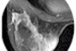
Left: contrast-enhanced T1-weighted image shows multiple rim-enhancing masses in both hemispheres. The MR features are indeterminate for abscess or tumor. Right: rCBV map shows regions of decreased blood volume corresponding to rim-enhancing lesions seen on image at left. The lesions proved to be nocardial infection.
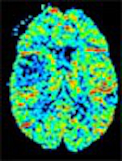
Left: Contrast-enhanced axial T1-weighted image shows multiple rim-enhancing masses in both hemispheres. The MR features are indeterminate for abscess or tumor. Right: rCBV map shows regions of increased cerebral blood volume (equal to that of normal cortex) corresponding to rim-enhancing mass on image at left. The lesion was found at surgery to be glioblastoma multiforme. All images courtesy of Dr. James Provenzale, Duke University Medical Center, Durham, NC.
According to the results, tumors had a much lower rCBV ratio (mean 1.40, range 1.03-2.20) than abscesses (mean 0.82, range 0.61-0.95), compared to normal white matter. The Wilcoxon/Kruskal-Wallis test of rank sums showed p=0.03.
"As you can see, there was no overlap between the populations," Provenzale said.
Potential study limitations include the need to use small ROI and labor-intensive measurements, as well as inexact correspondence of the corresponding T1 and rCBV maps due to patient motion, he said.
"Previous treatment of three of the tumors may have influenced the results. However, in our experience treatment has actually lowered blood volume rather than elevated it because there was a decrease in the vascularity of the lesion. Had the tumors not been treated, the difference (in ratios) might have been even more apparent."
Despite the small sample size, the results reached statistical significance, he said.
"In conclusion, rim-enhancing tumors could be distinguished based on hemodynamic characteristics," Provenzale said. "Further studies using a larger patient population are needed to confirm these preliminary results."
By Eric BarnesAuntMinnie.com staff writer
February 16, 2001
Copyright © 2001 AuntMinnie.com