In order to avoid missed breast lesions, functional MRI should be performed when the extraction-flow volume is at its lowest in a woman’s menstrual cycle, according to Dr. Eren Yeh from Massachusetts General Hospital in Boston. At the 2000 RSNA meeting, Yeh presented the results of a study that looked at the influence of the menstrual cycle on fMRI for suspected breast lesions.
"MR signal enhancement depends on several factors: the pathophysiology of the tissue and intrinsic MR properties of the tissue, including T1 signal intensity, relaxation time, and type of pulse sequence," Yeh explained.
In this patient population, 22 premenopausal women with a suspected or diagnosed unilateral breast lesion were examined with a Signa 1.5-tesla system (GE Medical Systems, Waukesha, WI). Bilateral MR was performed using gradient-echo and dynamic contrast-enhanced echo-planar imaging, with and without gadolinium-DTPA injection.

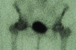
A 46-year-old female in days 7-3 of her menstrual cycle. Above is a high-resolution, gadolinium-enhanced MR image. Below is a functional map depicting the mean EF of the left breast (4 ml/100g/min) and a mean EF of the right breast (20 ml/100g/min). This is an example of how functional imaging supports scanning during the early part of the menstrual cycle when mean EF and signal enhancement of the breast tissue is at its lowest. Image courtesy of Dr. Eren Yeh.
The researchers measured precontrast T1 relaxation times of tissue in the non-suspect breast. Next, they measured changes in signal intensity after administering the contrast agent, and calculated extraction-flow product (EF) maps. EF reflects blood flow and capillary permeability, and is used to characterize breast tumors, which show an increased EF value because of angiogensis.
Finally, T1 and EF values in the control breast were recorded as a function of 5 menstrual stages: proliferative (days 7-3), follicular (days 8-14), luteal (days 20-15), secretory (days 27-21), and menstrual (days 28-2).
The T1 and EF results were as follows:
- Proliferative phase, T1 962 ms; EF 5.1 ml.100g-1.min-1
- Follicular phase, T1 1,092 ms; EF 8.1 ml.100g-1.min-1
- Luteal phase, T1 1,102 ms; EF 9.7 ml.100g-1.min-1
- Secretory phase, T1 1,157 ms; EF 13.4 ml.100g-1.min-1
- Menstrual phase, T1 1,158 ms; EF 8.1 ml.100g-1.min-1
"At days 7-3, the EF was the lowest at 5.5, and it was the highest at days 27-21 with an EF up to 14.6," Yeh said. "Signal enhancement also varied significantly throughout the menstrual cycle. For days 7-3, enhancement was lowest."
Since breast lesions are characterized by high EF values, fMRI should be performed when the EF of normal tissue is at its lowest, Yeh concluded.
In terms of T1 relaxation time, Yeh cited previous research by investigators at the University of Aberdeen in Scotland. In that paper, eight healthy women were imaged with MRI during 4-8 consecutive menstrual cycles. The total breast volume, water content, parenchymal volume, and T1 relaxation time were lowest between days 6 and 15. At days 28-16, T1 relaxation time rose by 15.1% (British Journal of Obstetrics and Gynecology, July 1990, Vol.97:7, pp.595-602).
By Shalmali PalAuntMinnie.com staff writer
February 14, 2001
Related Reading
Breast MRI's sensitivity poses problems for patient management, NCBC lecture shows, April 11, 2000
Click here to post your comments about this story. Please include the headline of the article in your message.
Copyright © 2001 AuntMinnie.com