F-18 FDG-PET measurements are comparable to those from cardiac MRI in breast cancer patients undergoing chemotherapy, researchers in Thailand have reported.
The finding is from a comparison between multiple PET acquisition techniques and cardiac MRI for measuring left ventricular ejection fraction (LVEF), a significant indicator of the severity of cardiac dysfunction, noted lead author Pariyaporn Toompong, MD, of the Chulabhorn Royal Academy in Bangkok, and colleagues.
"The use of PET-based methods to measure LVEF may be advantageous over cardiac MRI in terms of convenience and accessibility in patients with breast cancer," the authors wrote. The study was published March 17 in the Journal of Nuclear Medicine Technology.
Accurate LVEF monitoring is essential in breast cancer patients receiving cardiotoxic chemotherapy, where early detection of cardiac dysfunction can guide treatment decisions, the authors explained. While cardiac MRI is considered the gold standard in these cases, its use is constrained by long scanning times, repeated breath-holding requirements, and contraindications in patients with metallic implants, the authors noted.
Conversely, F-18 FDG-PET has shown promise in several recent studies, and in this prospective experiment, the researchers aimed to provide further evidence of its value. The group recruited 10 breast cancer patients (mean age 52.9 years old) presenting for LVEF assessment between July 2022 and February 2023.
All patients underwent two scans. First, a PET/CT scan and then, within 60 minutes, a PET/MRI scan (Biograph Vision 600 Edge, Siemens Healthineers), with the cardiac MRI measurements acquired simultaneously with the 3-tesla component of the hybrid scanner. The researchers reconstructed the PET images from the PET/CT scans using three techniques (dual-gated, AI-assisted dual-gated list-mode, and single-gated electrocardiography-gated) and then compared LVEF measurements across modalities.
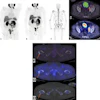
 Evaluation of gated PET data using Corridor4DM. Delineations of LV borders are shown as end-diastolic (ED) frame in horizontal long axis (HLA), as end-systolic (ES) frame in HLA, as ED frame in vertical long axis (VLA), and as ES frame in VLA. Time–volume curve of LV over different phases is shown with volumetric results.Journal of Nuclear Medicine Technology
In addition, dual-gated PET/CT demonstrated the smallest average percentage difference from cardiac MRI at 6.4%, followed by single-gated PET/CT (7.1%), list-mode PET/CT (7.4%), and PET/MRI (9.5%).
“F-18 FDG-PET–based methods may play an increasingly important role in noninvasive cardiac assessment, as they showed a good correlation with cardiac MRI for the evaluation of LV volume, LVEF, and myocardial wall motion in patients with breast cancer,” the researchers wrote.
While the study was limited by the small sample size, the findings support PET-based LVEF assessment as a clinically viable option in patients already undergoing F-18 FDG-PET for oncologic staging or response evaluation, the authors suggested. Larger prospective validation studies are warranted, they wrote.
“The use of PET-based methods to measure LVEF may be advantageous in terms of convenience and accessibility in this population,” the researchers concluded.
The full study is available here.