A recently developed PET camera appears to enable imaging during head motion and has revealed brain patterns in patients when they cough, according to a study published January 8 in Scientific Reports.
The study found that brain activity during coughing is more localized than previously believed, with the researchers noting that previous efforts using functional magnetic resonance imaging (fMRI) have been limited by artifacts caused by head movement.
“No real brain activity occurring during cough can be successfully captured with existing techniques. To address this limitation, we have recently developed a PET camera equipped with a free-moving function that enables imaging during head motion,” wrote lead author Takafumi Sugi, MD, of Hamamatsu City Rehabilitation Hospital in Shizuoka, Japan, and colleagues.
Coughing is a protective reflex that protects the airways from harmful substances and is necessary to maintain normal airway patency. This is difficult in patients with dysphasia, a neurologic condition that may be caused by stroke, the authors noted. They wrote that in their daily clinical practice, for instance, they promote voluntary coughing in dysphagic patients using a procedure called “cough‐inducing method with tartaric acid” (CiTA).
In this study, the researchers evaluated whether the experimental PET system could clarify brain activity during CiTA. This would allow clinicians to predict whether CiTA will be effective before attempting it in patients with brain lesions who have sustained damage to the regions related to cough control, they wrote.
The group enrolled 24 healthy patients who underwent separate O-15 water PET scans during four tasks: resting, voluntary cough, induced cough (where participants were induced by CiTA), and suppressed cough.
Participants were fitted with masks for cough measurements during two-minute PET scans. The tussive agent (tartaric acid) was delivered through the mask connected to a nebulizer via a nylon tube, with the nebulizer turned on for 20 seconds and off for 10 seconds for a total of two cycles.
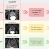
 Statistical parametric mapping (SPM) results. Brain regions with significant activation in voluntary cough (A), induced cough (B), and suppressed cough (C) compared with rest. Image courtesy of Scientific Reports.
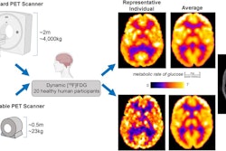
During the PET scans, the participants also wore a head cap with four LED markers. The locations of these markers were tracked by two high-speed cameras mounted at the back of the scanner’s gantry. This data was integrated by an algorithm during image reconstruction to correct for head motion every four milliseconds.
According to the results, whole-brain analyses revealed that volunteer cough chiefly activated the cerebellum extending to the pons. In contrast, CiTA-related tasks (induced cough and suppressed cough) activated the higher sensory regions of the cerebral cortex and associated brain regions.
“Using the current head motion correction method, we found that brain activity during the coughing task was much more localized than previously reported,” the authors wrote.
Significantly, the study identified the pons and brainstem as the most important regions in the control of stimulated cough and highlighted that the cerebellum is a key player in the regulation of the cough reflex, they added.
Ultimately, the study provides new basic insights on how the brain controls cough and shows promise for the use of the new PET scanner in clinical scenarios, the authors wrote.
“To confirm this, the current study is worth extending into a future study in dysphagic patients,” the group concluded.
The full study can be found here.