PET scans for the first time show brain activity that could potentially explain persistent depressive symptoms in patients after COVID-19 infections, according to a study published May 31 in JAMA Psychiatry.
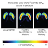
Researchers at the University of Toronto in Ontario, Canada, found evidence of brain gliosis -- an inflammatory response to injury -- on PET scans of mild to moderate COVID-19 participants with persistent depression and other cognitive symptoms compared to a healthy group. The findings offer a potential new direction for clinical care, the authors suggested.
"Depressive symptoms with or without other cognitive symptoms, after an acute episode of mild to moderate COVID-19 illness, hereinafter termed COVID-DC, is a major public health problem," wrote lead author Joeffre Braga, a doctoral student, and colleagues.
Persistent depressive and cognitive symptoms occur after COVID-19 infection in up to 15% of mild to moderate cases, depending on the variant, according to the authors. Research has shown that gliosis is associated with depressive symptoms in several neuropsychiatric diseases, and the authors hypothesized the mechanism may be involved in these symptoms in COVID-19 patients.
For brain imaging, they used PET imaging with a radiotracer (F-18 FEPPA) designed to measure translocator protein (TSPO), a protein overexpressed by activated glial cells, and which serves as a biomarker for neuroinflammation.
"This case-control study is, to our knowledge, the first study to assess brain gliosis in postacute sequelae of SARS-CoV-2 infection," the group wrote.
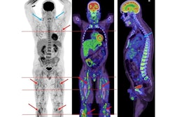
Between April 2021 to June 2022, the researchers performed PET scans in 40 participants between 18 and 72 years old at their hospital in Toronto. Twenty patients had been diagnosed with COVID-DC and 20 healthy participants served as controls. The main inclusion criterion for participants with COVID-DC was onset of a new major depressive episode (MDE) within three months.
The findings revealed increased expression of TSPO in patients with persistent neurocognitive symptoms after COVID-19 infection compared to healthy controls. Specifically, increases were most elevated in the ventral striatum (22%) and dorsal putamen (20%) -- regions of the brain that play crucial roles in movement, reward processing, and motivation.
"These findings suggest that gliosis, especially in the ventral striatum and dorsal putamen, may reflect injury, ongoing inflammation, or both and provide directions for further therapeutic development," the group wrote.
In an accompanying editorial, Dr. Alexander Gerhard, of the University of Manchester in England, noted that the work has important "pilot character" and that one could speculate that suppression of microglial activation might lead to improvement of these symptoms in COVID-19 patients.
However, future work aiming to understand the potential neuroinflammatory basis of cognitive symptoms and mood changes after COVID-19 infection is warranted, he added.
"While this is an important piece in the jigsaw puzzle of neuroinflammation in chronic neurological disease, it is important to keep in mind that we still lack understanding of the complex picture," Gerhard concluded.