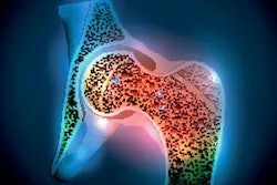
Dual-energy x-ray absorptiometry (DEXA) scans indicate that vitamin D supplementation during pregnancy results in greater bone mineral density in children up to age 7, according to a group in the U.K.
The finding is from a follow-up study of a large trial that demonstrated a similar positive effect of vitamin D supplementation in children at birth and at 4 years old compared with women who were not supplemented, noted lead author Rebecca Moon, PhD, of the University of Southampton, and colleagues.
“Demonstrating persistence of this effect is important to understanding whether maternal vitamin D supplementation could be a useful public health strategy to improving bone health,” the group wrote. The study was published September 19 in the American Journal of Clinical Nutrition.
A previous study conducted between 2008 and 2014 called the MAVIDOS trial tested the hypothesis that the offspring of pregnant women who took supplemental vitamin D may have higher bone mass. Compared with women who were not supplemented, initial results published in 2016 showed a positive bone mineral density (BMD) effect of 1,000 IU/day of vitamin D during pregnancy on children at birth and at 4 years old.
In this study, which included data from between November 2016 and April 2022, 447 children were followed up at the ages of 6 and 7. The children had DEXA scans of whole-body-less-head (WBLH) and lumbar spine, from which the researchers derived bone area (BA), bone mineral content (BMC), BMD, and bone mineral apparent density (BMAD).
The researchers used linear regression analysis to compare findings among the two groups (placebo and intervention groups), adjusting for age, sex, height, weight, duration of consumption of human milk, and vitamin D use.
According to the findings, gestational vitamin D supplementation resulted in higher WBLH BMC (0.15 standard deviation, or SD), BMD (0.18 SD), BMAD (0.18 SD), and lean mass (0.09 SD) compared with placebo. Interestingly, the effect of pregnancy vitamin D on bone outcomes was similar in children at ages 4, 6, and 7, the researchers noted.
“Supplementation with cholecalciferol 1000 IU/day during pregnancy resulted in greater offspring BMD and lean mass in mid-childhood versus placebo in this exploratory post-hoc analysis,” the group wrote.
The MAVIDOS study is the largest to date of pregnancy vitamin D supplementation to assess offspring BMD using DEXA and has the furthest duration of follow-up, but is not without limitations: Namely, due to an ethical stipulation, women who were very deficient in vitamin D and who would perhaps be expected to derive the greatest benefit from supplementation were excluded, the researchers noted.
While this limitation would be expected to favor the “null hypothesis,” the study nonetheless found a positive effect of vitamin D supplementation, they wrote.
“These findings suggest that pregnancy vitamin D supplementation may represent a population health strategy to improve bone health, although further work is needed to demonstrate persistence of this effect into adulthood,” the group concluded.
The full study is available here.