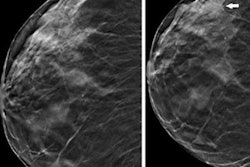
X-ray tomosynthesis shows benefits over conventional x-ray for lumbar spine imaging, not only producing high-quality images but also reducing radiation dose, according to a study published September 19 in Academic Radiology.
The findings may offer clinicians a better way to assess the spinal column, wrote a team led by Nora Conrads, MD, of the University Hospital Würzburg in Germany.
"In a growing proportion of elderly patients who present with spinal pathologies, reduced bone density impairs diagnostic assessment by means of standard x-ray imaging even further due to high radiolucency," Conrads and colleagues noted. "As a result, the increased risk of misreading lower spine pathologies on conventional radiographs has led to more requests for second-line imaging, which is associated with higher radiation exposure. With its superimposition-free visualization of vertebrae under physiological load, upright tomosynthesis may offset the diagnostic uncertainties inherent in radiography when used as a first-line imaging method."
Digital tomosynthesis generates image slices from low-dose projections at different angles, the team explained; it has been established as an effective technique in breast imaging and has begun to be explored for use in orthopedic surgery and musculoskeletal applications. X-ray tomosynthesis may be a "suitable alternative to multidetector [CT] of the appendicular skeleton for trauma or preoperative imaging," eliminating the problem of superimposition of anatomical structures in spinal x-ray imaging, the group wrote.
"While cross-sectional imaging by means of CT facilitates superimposition-free visualization of the osseous structures, it is associated with considerably increased radiation exposure compared to conventional radiography," Conrads and colleagues noted.
The team conducted a study that included eight cadavers and used a gantry-free twin robotic scanner to procure lateral conventional x-rays and x-ray tomosynthesis images of the lumbar spine. Five musculoskeletal radiologists assessed image quality between the two techniques, including the extent of any image quality deterioration near scan volume margins.
The study showed that x-ray tomosynthesis offered substantial dose reduction compared to conventional x-ray. Diagnostic image quality was highest using a 30 fps (frames per second) wide-angle tomosynthesis protocol. The team also reported good-to-excellent interrater reliability on x-ray tomosynthesis imaging (range, 0.85 to 0.95, with 1 as reference).
| Dose reduction for lumbar spine imaging comparing standard x-ray with x-ray tomosynthesis | |||
|---|---|---|---|
| Measure | Standard x-ray | X-ray tomosynthesis | p-value |
| Radiation dose | 77.7 dGy*cm2 | 3.8 dGy*cm2 | ≤ 0.021 |
"Combining minimal radiation dose with superimposition-free visualization, 30 fps wide-angle tomosynthesis superseded radiography in all evaluated aspects," the authors concluded.
The complete study can be found here.


