Dynamic chest radiography appears promising for evaluating responses in cystic fibrosis (CF) patients after they receive treatment for lung flare-ups, according to a study published March 15 in Radiology.
In a prospective pilot study, a U.K. group evaluated whether dynamic chest radiography could be used to measure the effects of treatment in cystic fibrosis patients experiencing pulmonary exacerbations. The technique showed improvements in patients' diaphragm speed and range of chest wall movement after treatment, the authors wrote.
"Dynamic chest radiography shows potential as a rapid and useful imaging tool to assess the effect of pulmonary exacerbations in cystic fibrosis," wrote first author Dr. Thomas FitzMaurice, PhD, a research fellow at Liverpool Heart and Chest Hospital.
Cystic fibrosis is a progressive, genetic disease that causes sticky, thick mucus to build up in organs, primarily the lungs. This makes it difficult for patients to breathe. Treatments include methods to clear the lungs, including the use of anti-inflammatory drugs.
Traditionally, simple spirometry is used to assess whether treatments are working in patients who experience pulmonary exacerbations, or flare-ups, but spirometry requires repeated forced breathing maneuvers and the use of a mouthpiece and nose clip that interfere with airflow, according to the authors. The test may be difficult to perform in some individuals, they added.
Dynamic chest radiography is a novel real-time digital fluoroscopic imaging system that produces clear, wide field-of-view diagnostic images of the thorax and diaphragm in motion, alongside novel metrics on moving structures within the thoracic cavity.
Previously, FitzMaurice and colleagues showed that the technique may be useful for visualizing diaphragm motion in a series of patients with suspected damage of the phrenic nerve, which controls the diaphragm and, ultimately, normal breathing.
In this study, the group enrolled 20 adult patients with cystic fibrosis who subsequently underwent dynamic chest radiography before and after treatment for pulmonary exacerbations between December 2019 and August 2020.
Dynamic chest radiography images were acquired over 10 seconds with patients in the erect position using a system from Konica Minolta Healthcare. Imaging was performed within 48 hours of pulmonary exacerbation diagnosis and again within 48 hours after the patients' flare-ups had resolved.
The researchers analyzed metrics for measuring improvements after treatment. Dynamic chest radiography measured projected lung area, hemidiaphragm midpoint position, and speed during tidal and deep breathing.
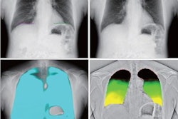
 Still image from a video published online showing a dynamic chest radiograph acquired during tidal and deep breathing. Image courtesy of Radiology.
Following treatment for pulmonary exacerbation, dynamic chest radiography showed an improvement in the depth of median right (18 mm to 25 mm) and left (13 mm to 19 mm) hemidiaphragm excursion during deep breathing.
In addition, the researchers observed an improvement in left (7 mm/sec to 11 mm/sec) and right (6 mm/sec to 9 mm/sec) mean expiratory diaphragm velocity and improvements in tidal breathing excursion of both median left (12 mm to 15 mm) and right (11 mm to 13 mm) hemidiaphragms after treatment.
"Dynamic chest radiography demonstrated improvement in diaphragm speed and range of chest wall movement during respiration after treatment for pulmonary exacerbations in cystic fibrosis," the authors stated.
The study provides insights into pulmonary dynamics and suggests that dynamic chest radiography may be of particular use in individuals in whom spirometry is difficult to perform, the authors suggested.
Dynamic chest radiography also provides the ability to perform imaging away from attending clinicians, which could reduce exposure to potential aerosol generation by patients with transmissible respiratory infections, they added.
Ultimately, the technique warrants further investigation, FitzMaurice and colleagues concluded.