Dear Digital X-Ray Insider,
One of the great things about digital radiography (DR) is the ability to manipulate images in ways you can't with analog x-ray -- panning, zooming, rotating, and adding annotations and measurements. But could you unknowingly be degrading your images in the process?
The problem is due to a phenomenon called bit reduction quantization, which occurs when images acquired at higher bit rates are reduced to lower bit rates during image manipulation. Quantization can occur any time you alter and save changes to an original 14-bit DR image using 12-bit PACS software.
Researchers from the Mayo Clinic in Rochester, MN, investigated whether quantization affected the image quality of DR and MR images, and their work is the subject of this edition's Insider Exclusive. Click here to learn more about what they found.
In other news in the world of DR, associate editor Cynthia E. Keen contributes an article describing how computer-aided detection software was able to find overlooked lung lesions on DR and computed radiography studies.
Also learn about new guidelines from the Society of Interventional Radiology on patient radiation dose during fluoroscopy procedures.
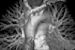
And finally, find out how radiographic findings on small-bowel exams can help find internal hernia following Roux-en-Y gastric bypass procedures for morbidly obese patients.
If you have tips or ideas on topics you'd like to see covered in the Digital X-Ray Community, drop me a line at [email protected].