
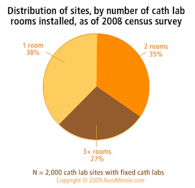
An estimated total of 4,225 fixed cath lab rooms are installed in 2,000 sites nationwide. The average number of cath lab rooms per site is 2.1 rooms. As of this 2008 census survey, 38% of all cath lab sites had one room, 35% had two rooms, and 27% had three or more rooms.

Based on responses to IMV’s 2008 Cardiac Catheterization Lab Survey of U.S. Hospitals and Nonhospitals.
AuntMinnie's IMV MarketStat is provided to AuntMinnie.com by IMV Medical Information Division, Inc. of Des Plaines, IL.

Click here to buy complete IMV Market Reports
MarketStat ArchivesHard-copy film use at sites with digital mammography units
Nuclear medicine -- Average waiting time
Radiation oncology -- Types of images used in radiation therapy treatment plans
Vascular MRI -- Percent of sites performing and percent of MRI procedures
Single versus multislice detectors in CT installed base
Budgets for nuclear medicine radiopharmaceuticals
Formats used for images sent or received by radiation oncology departments
MRI adult versus pediatric patient visit mix
Cath lab device budgets for 2006
Copyright © 2009 AuntMinnie.com
![Representative example of a 16-year-old male patient with underlying X-linked adrenoleukodystrophy. (A, B) Paired anteroposterior (AP) chest radiograph and dual-energy x-ray absorptiometry (DXA) report shows lumbar spine (L1 through L4) areal bone mineral density (BMD). The DXA report was reformatted for anonymization and improved readability. The patient had low BMD (Z score ≤ −2.0). (C) Model (chest radiography [CXR]–BMD) output shows the predicted raw BMD and Z score in comparison with the DXA reference standard, together with interpretability analyses using Shapley additive explanations (SHAP) and gradient-weighted class activation maps. The patient was classified as having low BMD, consistent with the reference standard. AM = age-matched, DEXA = dual-energy x-ray absorptiometry, RM2 = room 2, SNUH = Seoul National University Hospital, YA = young adult.](https://img.auntminnie.com/mindful/smg/workspaces/default/uploads/2026/04/ai-children-bone-density.0snnf2EJjr.jpg?auto=format%2Ccompress&fit=crop&h=100&q=70&w=100)


![Representative example of a 16-year-old male patient with underlying X-linked adrenoleukodystrophy. (A, B) Paired anteroposterior (AP) chest radiograph and dual-energy x-ray absorptiometry (DXA) report shows lumbar spine (L1 through L4) areal bone mineral density (BMD). The DXA report was reformatted for anonymization and improved readability. The patient had low BMD (Z score ≤ −2.0). (C) Model (chest radiography [CXR]–BMD) output shows the predicted raw BMD and Z score in comparison with the DXA reference standard, together with interpretability analyses using Shapley additive explanations (SHAP) and gradient-weighted class activation maps. The patient was classified as having low BMD, consistent with the reference standard. AM = age-matched, DEXA = dual-energy x-ray absorptiometry, RM2 = room 2, SNUH = Seoul National University Hospital, YA = young adult.](https://img.auntminnie.com/mindful/smg/workspaces/default/uploads/2026/04/ai-children-bone-density.0snnf2EJjr.jpg?auto=format%2Ccompress&fit=crop&h=112&q=70&w=112)


