Dear AuntMinnie Member,
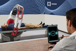
While AI models have been trained to estimate bone mineral density from adult chest x-rays, no model to date has been assessed for use in children, according to a group in Seoul, South Korea. To that end, they developed a deep learning model for the task and suggest its performance supports translation into practice. Read the details here.
After you’ve read that story, check out our interview with the lead author of an article in Radiology who suggested that radiologists are not able to easily distinguish AI-generated “deepfake” x-ray images from authentic ones.
Here are a few other stories on AI x-ray research, which illustrate the value and limitations of the technology:
Aortic calcification visible on chest x-rays is associated with poorer overall survival in patients following minor lower extremity amputation, according to a team in Finland.
Researchers in Ethiopia developed a new strategy for diagnosing tuberculosis in low-resource, high-incidence settings by photographing film x-rays to create digital files and then feeding them to AI.
A group in the U.K. found that automatically flagging suspicious chest X-rays with AI for urgent radiologist review did not reduce time to CT or lung cancer diagnosis.
Neither radiologists nor an AI algorithm can consistently identify interstitial lung abnormalities on standard chest x-rays, according to a study published in the American Journal of Roentgenology.
We've also posted a few articles on research that delved into the value of x-rays. Researchers found that young athletes with low back pain may not need to undergo advanced imaging beyond initial x-rays (and yet they do). Meanwhile, a team at the University of Michigan found that plain x-ray remains commonly used in initial imaging of adults with maxillofacial trauma, despite clear guidelines favoring CT.
Finally, we covered a dual-energy x-ray absorptiometry study by researchers at the University of North Carolina at Chapel Hill that found exposure to “forever chemicals” during childhood is associated with lower bone mineral density (BMD) in early adolescence.
For more x-ray news, be sure to check in regularly with our Digital X-Ray content area. And as always, if you have x-ray topics you'd like us to consider, please contact me.
Will Morton
Associate Editor
AuntMinnie.com


![Representative example of a 16-year-old male patient with underlying X-linked adrenoleukodystrophy. (A, B) Paired anteroposterior (AP) chest radiograph and dual-energy x-ray absorptiometry (DXA) report shows lumbar spine (L1 through L4) areal bone mineral density (BMD). The DXA report was reformatted for anonymization and improved readability. The patient had low BMD (Z score ≤ −2.0). (C) Model (chest radiography [CXR]–BMD) output shows the predicted raw BMD and Z score in comparison with the DXA reference standard, together with interpretability analyses using Shapley additive explanations (SHAP) and gradient-weighted class activation maps. The patient was classified as having low BMD, consistent with the reference standard. AM = age-matched, DEXA = dual-energy x-ray absorptiometry, RM2 = room 2, SNUH = Seoul National University Hospital, YA = young adult.](https://img.auntminnie.com/mindful/smg/workspaces/default/uploads/2026/04/ai-children-bone-density.0snnf2EJjr.jpg?auto=format%2Ccompress&fit=crop&h=167&q=70&w=250)