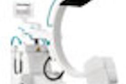
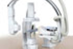
(Booth 2554) In its RSNA booth, Konica Minolta Medical Imaging USA will highlight its Regius Nano CR computed radiography unit and Regius 370 Upright DR digital radiography system.
Regius Nano CR is a single-bay system, designed for low-volume facilities and ancillary departments. It includes a cassette release handle for easy removal of a trapped cassette, an optics sweeper that allows a user to easily clean the optical unit, and Konica Minolta's rigid image plate technology. Regius Nano CR is available in two models: Enterprise Solution for hospitals and healthcare systems and Clinic Solution for clinics and physician offices.
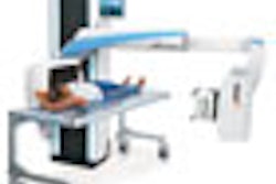
Regius 370 Upright DR features a cesium bromide storage phosphor detector and is compatible with any of a facility's existing x-ray generators or tubes, according to the Wayne, NJ, company. It can scan an image at a rate of 210 exposures an hour.
Konica Minolta also plans to display its DryPro 832 medical laser film imager, which can produce 90 14 x 17-inch films per hour.

![Representative example of a 16-year-old male patient with underlying X-linked adrenoleukodystrophy. (A, B) Paired anteroposterior (AP) chest radiograph and dual-energy x-ray absorptiometry (DXA) report shows lumbar spine (L1 through L4) areal bone mineral density (BMD). The DXA report was reformatted for anonymization and improved readability. The patient had low BMD (Z score ≤ −2.0). (C) Model (chest radiography [CXR]–BMD) output shows the predicted raw BMD and Z score in comparison with the DXA reference standard, together with interpretability analyses using Shapley additive explanations (SHAP) and gradient-weighted class activation maps. The patient was classified as having low BMD, consistent with the reference standard. AM = age-matched, DEXA = dual-energy x-ray absorptiometry, RM2 = room 2, SNUH = Seoul National University Hospital, YA = young adult.](https://img.auntminnie.com/mindful/smg/workspaces/default/uploads/2026/04/ai-children-bone-density.0snnf2EJjr.jpg?auto=format%2Ccompress&fit=crop&h=100&q=70&w=100)


![Representative example of a 16-year-old male patient with underlying X-linked adrenoleukodystrophy. (A, B) Paired anteroposterior (AP) chest radiograph and dual-energy x-ray absorptiometry (DXA) report shows lumbar spine (L1 through L4) areal bone mineral density (BMD). The DXA report was reformatted for anonymization and improved readability. The patient had low BMD (Z score ≤ −2.0). (C) Model (chest radiography [CXR]–BMD) output shows the predicted raw BMD and Z score in comparison with the DXA reference standard, together with interpretability analyses using Shapley additive explanations (SHAP) and gradient-weighted class activation maps. The patient was classified as having low BMD, consistent with the reference standard. AM = age-matched, DEXA = dual-energy x-ray absorptiometry, RM2 = room 2, SNUH = Seoul National University Hospital, YA = young adult.](https://img.auntminnie.com/mindful/smg/workspaces/default/uploads/2026/04/ai-children-bone-density.0snnf2EJjr.jpg?auto=format%2Ccompress&fit=crop&h=112&q=70&w=112)


