Chest CT scans can predict future thoracic vertebral bone mineral density loss by measuring paraspinal muscle attenuation, a finding that allows routine imaging to serve as an opportunistic screening tool for musculoskeletal risk without requiring additional scans.
- Paraspinal muscle attenuation independently predicts thoracic vertebral bone mineral density loss from routine noncontrast chest CT scans
- Study of 1,316 adults over 6 years showed muscle attenuation improved BMD prediction accuracy by 7% and fracture risk prediction from 0.76 to 0.82 AUC
- Participants with lowest muscle attenuation had significantly lower baseline bone density (176 vs 205 mg/cm³) and were older, predominantly female, and White
- Deep learning algorithms automatically segment paraspinal muscles and measure attenuation in Hounsfield units, transforming labor-intensive assessment into practical opportunistic imaging
- Chest CT is well-suited for musculoskeletal risk assessment as it quantifies muscle, fat, and bone biomarkers without additional imaging acquisition
Chest CT scans can predict future thoracic vertebral bone mineral density (BMD) loss by measuring paraspinal muscle attenuation, according to a study published September 15 in Radiology.
The findings suggesting that the approach could extend the diagnostic reach of routine chest CT into musculoskeletal risk stratification, noted lead author Quincy Hathaway, MD, PhD, of the University of Pennsylvania, and colleagues.
“Paraspinal muscle attenuation determined from noncontrast chest CT represents an opportunistic imaging biomarker that provides independent and incremental value for predicting longitudinal thoracic vertebral bone mineral density (BMD) loss,” the group wrote.
Muscle and bone loss commonly coexist with aging, but potential links between paraspinal muscle – a group that runs vertically along both sides of the spinal column from the base of the skull to the pelvis – and thoracic vertebral BMD loss remain poorly understood, the authors wrote.
To address the gap, the researchers drew on data from the Multi-Ethnic Study of Atherosclerosis (MESA), a prospective, population-based U.S. cohort of nearly 7,000 adults aged 45 to 84 years at six centers. They analyzed noncontrast chest CT scans from 1,316 participants (mean age, 67.2 years old; 694 female) at baseline (2010–2012) and follow-up (2016–2018) over a 6-year period.
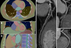
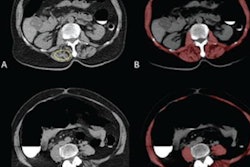
The researchers used previously developed deep learning algorithms to segment paraspinal muscles at vertebral levels T1 through T10 and to measure muscle attenuation in Hounsfield units from the baseline chest CT scans. They also applied a validated deep learning model to predict BMD from the same scans.
Key findings included the following:
Participants with the lowest paraspinal muscle attenuation had lower baseline thoracic vertebral BMD compared with participants in quartiles 2 to 4 (176 mg/cm3 vs 205 mg/cm3; P < .001) and were older, White, and predominantly female.
Paraspinal muscle attenuation increased the adjusted R2 value from 0.20 to 0.27 for follow-up BMD prediction (7% absolute increase).
Paraspinal muscle attenuation improved prediction of incident vertebral fracture beyond BMD alone (area under the receiver operating characteristic curve, 0.82 versus 0.76; p = .02).
“Noncontrast chest CT is well suited for opportunistic musculoskeletal risk assessment because it can help quantify muscle, fat, and bone biomarkers without additional imaging acquisition,” the group wrote.
Further studies are needed to determine whether paraspinal muscle attenuation is purely a biologic biomarker and provides independent predictive value and whether fracture prediction is reproducible in a larger prospective cohort, the group concluded.
In an accompanying editorial, Pranjal Rai, MD, a nuclear medicine fellow at the Mayo Clinic in Rochester, MN, and Amit Kumar Janu, MD, of the Advanced Centre for Treatment, Research and Education in Cancer in Navi Mumbai, India, wrote that the automated segmentation tools used in the study may help transform quantitative assessment of paraspinal muscle quality from a labor-intensive task into a practical opportunistic quantitative imaging measure that could be incorporated into routine interpretation of thousands of chest CT examinations.
However, as opportunistic imaging continues to evolve, successful clinical implementation will require more than accurate segmentation, they noted.
“Consensus regarding the optimal muscle groups, quantitative metrics, and clinically meaningful thresholds will be essential before these markers can be routinely incorporated into clinical workflows,” Rai and Janu wrote.
The full study is available here.