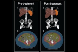
CT-derived body composition biomarkers show promise for categorizing the level of frailty in older adults, a study published July 20 in Academic Radiology has found.
The results underscore CT's role in effective assessment of frailty – a condition that can decrease individuals' quality of life, wrote a group led by Paul Bunch, MD, of Wake Forest University School of Medicine in Winston-Salem, NC.
"[There] is a need for better frailty screening tools, and our findings suggest that the CT-derived measures of skeletal muscle density and visceral adipose tissue may be useful," it noted.
As adults age, their level of frailty (which can include low grip strength, slow walking speed, and low skeletal muscle area and density) must be tracked so that effective preventive care can be offered, the team explained. CT-derived measurements of muscle and fat tissue, collected opportunistically and used for body composition analysis, could help in this regard, the team noted.
"Frail older adults are at increased risk for falls, hospitalizations, nursing home admissions, and mortality, and they report higher rates of loneliness and impaired quality of life," the group wrote. "Because the prevalence of frailty is expected to rise as the population ages, there is a growing need for prevention and treatment interventions."
To this end, the authors conducted a study to assess whether these CT measurements are associated with frailty, which it defined using an "electronic frailty index" applied to data from patients' health records. The index is scored in the following manner: fit (a score below 0.12), mildly frail (0.12 to 0.24), moderately frail (0.24 to 0.36), or severely frail (0.36 and above); the investigators used it to assess muscle metrics from CT images at the L3 vertebra level such as skeletal muscle area, skeletal muscle density, and intermuscular adipose tissue. The group also tracked patients' fat tissue metrics such as amounts of visceral adipose tissue and subcutaneous adipose tissue.
The study included 886 patients who underwent contrast-enhanced abdominopelvic CT. Of these, 382 (43%) met the "prefrailty" criteria and 138 (16%) were categorized as frail.
The investigators found the following:
- In men, mean skeletal muscle density was lower among pre-frail and frail men (p = 0.02 and p = 0.01, respectively) compared with healthy counterparts.
- No significant difference in skeletal muscle density were found among prefrail, frail, and healthy women.
- No significant skeletal muscle area, intermuscular adipose tissue, visceral adipose tissue, or subcutaneous adipose tissue were associated with frailty in either men or women.
The bottom line? CT-derived measures could help clinicians evaluate the level of frailty among their older adult patients -- and thus better strategize their care.
"Future studies are warranted to determine whether the application of radiomic analyses to CT-derived muscle and fat compartments could improve the assessment of frailty ... eventually, CT images may be leveraged to provide an individualized frailty assessment and serve not only as a means for diagnosing and risk-stratifying but also for targeting therapeutics and assessing treatment response," the study authors concluded.
The complete study can be found here.


