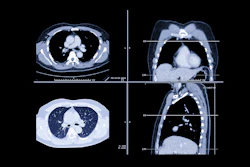
The combination of high-resolution CT and histopathological data boosts radiologists' ability to diagnose interstitial lung disease (ILD), a study published August 23 in General Thoracic and Cardiovascular Surgery has found.
The results suggest that using the combination of CT and clinical data could help clinicians better tailor treatment for patients with ILD who then develop lung cancer, according to a group led by Dr. Yoko Azuma of Toho University in Tokyo.
"An accurate diagnosis of [interstitial lung disease subtypes] according to both high-resolution CT scan findings and histological patterns is important for providing an appropriate treatment among patients with lung cancer who present with clinical interstitial lung disease," Azuma and colleagues wrote.
Interstitial lung disease can exhibit in several ways, each of which has a different pathophysiology, the investigators explained. One of these is idiopathic interstitial pneumonia (IIP), which then has a common subtype of idiopathic pulmonary fibrosis (IPF) that tends to correlate with poor patient outcomes.
Individuals with interstitial lung disease have higher rates of lung cancer -- as well as higher rates of morbidity as the result of lung cancer treatment -- and are also at risk of complications that develop within a month after surgical treatment.
Azuma and colleagues explored whether identifying the subtype of interstitial lung disease using CT and histopathogical data (the latter gained by using a microscope to examine tissue or cells) could help predict a patient's outcomes after lung cancer surgery. The team compared CT findings to histopathological data; histopathological characteristics included presence or absence of the following:
- A fibrous interstitium
- Emphysematous change (i.e., obstructed transfer of oxygen and carbon dioxide among the alveoli)
- Bronchiolocentric location (inflamed fibrotic areas in the small airways of the lungs)
- Absence of fibroblastic foci (defined as areas that consist of fibroblasts, myofibroblasts, and collagen)
The study included 104 patients diagnosed with ILD who also underwent lung resection surgery between January 2004 and December 2020. Patients were classified into four groups: those with IPF, those likely to have it, those for whom the presence of IPF was unclear, and those who did not have it.
The investigators found that the presence of idiopathic pulmonary fibrosis identified by CT and/or histopathologic results did have a prognostic effect. Patients with the presence of both had poorer five-year lung cancer survival rates: 22.8% compared with 67.9% in those who did not have the condition (p = 0.01). The researchers attributed this poorer survival rate in part to the fact that people with idiopathic pulmonary fibrosis have a higher incidence of postsurgical complications, ranging from 10.7% to 23.1%.
Subclassification of idiopathic interstitial pneumonia is an independent, predictive factor for overall survival in people with lung cancer, according to the authors.
"Our data showed that an accurate classification of interstitial lung disease and idiopathic interstitial pneumonias affects prognosis after lung cancer surgery," they concluded.