MRI myelography offers a less invasive way to detect cerebrospinal fluid (CSF) leaks in patients with spontaneous intracranial hypotension compared to the CT version of the same exam -- and performs comparably, according to a research letter published August 30 in JAMA Neurology.
The results could help clinicians expand their toolkit for patients suffering from this condition, wrote a team of researchers led by Dr. Angelique Sao-Mai Sy Tay of Cedars-Sinai Medical Center in Los Angeles.
"Eliminating radiation exposure, obviating the need for lumbar puncture and intrathecal contrast injection, and decreasing resource utilization and cost are among the most important advantages of MRM [MRI myelography] over CTM [CT myelography]," the researchers wrote.
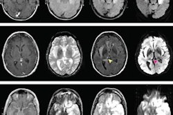
People with spontaneous intracranial hypotension are typically evaluated using CT myelography. But CT imparts radiation, and researchers have been exploring whether MRI myelography could offer a noninvasive alternative. Tay's team compared the diagnostic accuracy of CT myelography to MRI myelography for identifying CSF leaks in patients with spontaneous intracranial hypotension.
The study included 576 patients who underwent MR myelography and CT myelography exams. Of these, 276 (47.9%) were diagnosed with CSF leaks on both CT and MR myelography.
The investigators found that the MR myelography and CT myelography findings conflicted in less than 2% of patients with spontaneous intracranial hypotension. The group also found that positive agreement between the two exams was 99.6%, negative agreement was 97.7%, and overall agreement 98.6%.
The results indicate MRI myelography could be a feasible alternative to CT.
"MRM may be a suitable alternative to CTM for the initial evaluation of patients with spontaneous intracranial hypotension," the authors concluded.