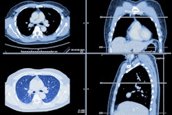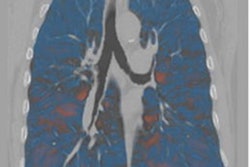
The pattern of parenchymal emphysema on baseline CT scans forecasts progression of the disease in patients who are current or former smokers both with and without chronic obstructive pulmonary disease (COPD), according to a study published online December 15 in Radiology.
The study results offer clinicians a framework with which they can track patient outcomes and even improve emphysema treatment, wrote a team led by Dr. Bilal El Kaddouri of Hospital Erasme, Université Libre de Bruxelles, in Brussels, Belgium.
"The correlation between visual emphysema patterns and subsequent progression of disease may provide a way to enrich a study population for treatment trials of emphysema," the group wrote.
CT is used in COPD patients to evaluate the presence, pattern, and severity of emphysema, and quantitative CT is also used to visualize COPD features and assess the effectiveness of emphysema treatment, El Kaddouri and colleagues noted. But there is little clinical research about the connection between visually assessed emphysema severity and disease progression, according to the researchers.
"We hypothesized that the presence of confluent or advanced destructive emphysema would be associated with more rapid progression of emphysema and gas trapping," they wrote.
El Kaddouri's group conducted a study that included 4,166 current and former smokers with and without COPD who participated in the Genetic Epidemiology of COPD (COPDGene) study between 2008 and 2011. The team assessed each person's Fleischner Society visual CT scores as well as quantitative inspiratory and expiratory scores, both at baseline and at five-year follow-up exams.
The investigators found that smokers with visible parenchymal emphysema at baseline CT showed marked disease progression at five years, even if the emphysema was mild or moderate, while those without visible disease did not.
| Change in lung density from baseline CT to 5-year follow up | ||
| Emphysema grade at baseline CT | Patients without COPD | Patients with COPD |
| Trace | -1.8 g/L | -2.4 g/L |
| Mild | -3.6 g/L | -4.6 g/L |
| Moderate | -3.1 g/L | -6.7 g/L |
| Confluent (advanced) | -4.0 g/L | -6.4 g/L |
The study indicates that the presence of emphysema at baseline CT is a good predictor of the disease's progression -- and that the Fleischner categories are useful, the authors concluded.
"The presence of visual parenchymal or paraseptal emphysema at CT in current or former smokers is an important predictor of subsequent progression of emphysema," they wrote. "The Fleischner Society emphysema grading system may be useful as a simple prognostic marker in smokers, with or without [COPD] and may have value in therapeutic trials and in clinical practice."