Complications from electronic cigarette (e-cigarette) or vaping product use-associated lung injury (EVALI) persist for months after patients treated in the hospital are discharged, according to a study published June 13 in the journal Chest.
The findings address a knowledge gap about the long-term effects of EVALI, wrote a team led by Dr. Rachel Eddy of the University of Ontario in London, Ontario, Canada.
"While nearly 3,000 e-cigarette-related hospitalizations have been reported in North America, the long-term outcomes in these patients have not been described," the team wrote.
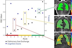
Eddy and colleagues followed an 18-year-old male patient who survived acute respiratory failure related to five months of e-cigarette use. The group found abnormal ventilation heterogeneity in the patient on hyperpolarized xenon-129 (Xe-129) gas with MRI months after he was discharged from the hospital, despite improvement in chest CT findings. His lung clearance index and oscillometry measures were also abnormal after this time period, the group noted.
"Our findings underscore the long-term functional impacts of e-cigarette-related lung injury in survivors of critical illness; longitudinal evaluations may shed light on the pathophysiologic mechanisms that drive e-cigarette-related lung disease," Eddy and colleagues concluded.