New CT and MR images of a patient in Michigan with COVID-19 appear to show the impact on the brain of "cytokine storm syndrome," when the body's immune system produces a flood of immune cells that can damage organs. The pictorial report was published online March 31 in Radiology.
Cytokine storm syndrome occurs when immune cells are overproduced by the body to fight an infection or cancer. The process can damage organs and can even be fatal. And based on literature from China, it has occurred in some patients with COVID-19.
In the new study, researchers from Henry Ford Health System in Detroit describe the first apparent case of COVID-19-associated acute necrotizing hemorrhagic encephalopathy, which they believe may have occurred due to cytokine storm syndrome.
The study patient is a female airline worker in her late 50s who presented at Henry Ford with a three-day history of cough, fever, and altered mental state in the form of confusion, lethargy, and disorientation, wrote a team led by Dr. Neo Poyiadji of Henry Ford. She was diagnosed as having been infected with SARS-CoV-2 via nasal swab and RT-PCT assay; she was also tested for herpes simplex virus 1 and 2, varicella-zoster virus, and West Nile virus, all of which were negative. She remains hospitalized and in serious condition, corresponding author Dr. Brent Griffith told AuntMinnie.com.
CT and MRI imaging showed the following:
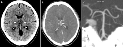
- Head CT images showed symmetric hypoattenuation within the bilateral medial thalami with a normal CT angiogram and CT venogram.
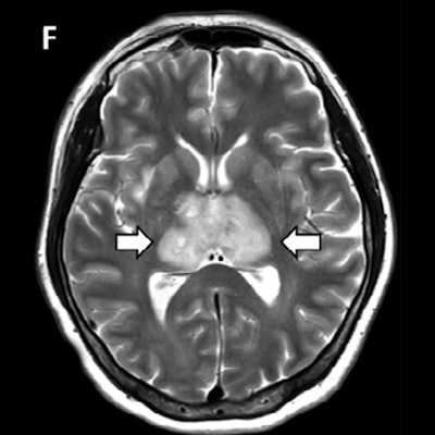
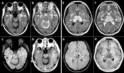
- Brain MRI images showed T2-weighted and fluid-attenuated inversion recovery (FLAIR) hyperintensity within the bilateral thalami, medial temporal lobes, and subinsular regions with areas of hemorrhage, as well as rim enhancement on postcontrast images.
Although rare, acute necrotizing encephalopathy is a complication of influenza and other viral infections. It's associated with the overproduction of immune cells and their compounds during cytokine storms, which results in blood-brain barrier breakdown, but without direct viral invasion or damage to nerve myelin, the researchers wrote.
 A, Image from noncontrast head CT demonstrates symmetric hypoattenuation within the bilateral medial thalami (arrows). B, Axial CT venogram demonstrates patency of the cerebral venous vasculature, including the internal cerebral veins (arrows). C, Coronal reformat of a CT angiogram demonstrates normal appearance of the basilar artery and proximal posterior cerebral arteries. All images courtesy of the RSNA.
 MRI images demonstrate T2 FLAIR hyperintensity within the bilateral medial temporal lobes and thalami (A, B, E, F) with evidence of hemorrhage indicated by hypointense signal intensity on susceptibility-weighted images (C, G) and rim enhancement on postcontrast images (D, H).
"Accumulating evidence suggests that a subgroup of patients with severe COVID-19 might have a cytokine storm syndrome," the team wrote. "The most characteristic imaging feature includes symmetric, multifocal lesions with invariable thalamic involvement. Other commonly involved locations include the brain stem, cerebral white matter, and cerebellum."
The authors hope the study will help their radiology peers recognize this rare condition as they deal with COVID-19.
"This is the first reported case of COVID-19-associated necrotizing hemorrhagic encephalopathy," the group wrote. "As the number of patients with COVID-19 increases worldwide, clinicians and radiologists should be watching for [this condition] among patients presenting with COVID-19 and altered mental status."


