
Preclinical imaging developer MILabs will install its in vivo optical imaging and diagnostic x-ray CT systems at the University of Lausanne and the Lausanne University Hospital in Switzerland.
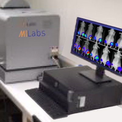
The systems will be installed at the In-Vivo Imaging Facility and serve a dual optical imaging function. The Duet Optical Imaging Module can be used on a docking station for 2D in vivo planar bioluminescence and fluorescence imaging of up to 10 mice at one time, the company said. Also, it can be mounted on the MILabs x-ray CT platform to allow for quantitative CT-guided 3D optical tomography images to be obtained with both bioluminescence and fluorescence probes.
MILabs' Duet Optical Imaging Module. Image courtesy of MILabs.


![Images show the pectoralis muscles of a healthy male individual who never smoked (age, 66 years; height, 178 cm; body mass index [BMI, calculated as weight in kilograms divided by height in meters squared], 28.4; number of cigarette pack-years, 0; forced expiratory volume in 1 second [FEV1], 97.6% predicted; FEV1: forced vital capacity [FVC] ratio, 0.71; pectoralis muscle area [PMA], 59.4 cm2; pectoralis muscle volume [PMV], 764 cm3) and a male individual with a smoking history and chronic obstructive pulmonary disorder (COPD) (age, 66 years; height, 178 cm; BMI, 27.5; number of cigarette pack-years, 43.2, FEV1, 48% predicted; FEV1:FVC, 0.56; PMA, 35 cm2; PMV, 480.8 cm3) from the Canadian Cohort Obstructive Lung Disease (i.e., CanCOLD) study. The CT image is shown in the axial plane. The PMV is automatically extracted using the developed deep learning model and overlayed onto the lungs for visual clarity.](https://img.auntminnie.com/mindful/smg/workspaces/default/uploads/2026/03/genkin.25LqljVF0y.jpg?auto=format%2Ccompress&crop=focalpoint&fit=crop&h=112&q=70&w=112)

