Tuesday, December 3 | 3:50 p.m.-4:00 p.m. | SSJ05-06 | Room S102CD
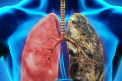
An artificial intelligence (AI) algorithm could help radiologists improve their diagnostic accuracy on CT lung cancer screening exams, according to this presentation.Although numerous scientific papers have shown that image texture features derived from computer-aided diagnosis (CAD) methods are associated with lung cancer, CAD has largely not been adopted into clinical practice for lung cancer screening. That's mostly because it's not clear that CAD can provide additive information beyond the radiographic characteristics used routinely by radiologists during interpretation, according to Peng Huang, PhD, of Johns Hopkins University in Baltimore.
"Since a direct comparison in diagnostic accuracy between CAD and radiologist reading is lacking, we conducted a matched case-control study to evaluate the added value of CAD in cancer diagnosis using CT images acquired right before lung biopsy or surgery," Huang told AuntMinnie.com.
To facilitate the comparison, they developed a machine-learning algorithm that was designed to mimic how a radiologist reads CT studies. It also extracts CT image texture features -- including radiomics features -- from the intra-, peri-, and extranodular regions, as well as the lymph nodes and other surrounding lung tissues.
The researchers then compared the diagnostic accuracy of the algorithm with that of three experienced radiologists on the same set of images. The machine-learning algorithm yielded higher accuracy, likely due to the independent information gained from its analysis of image texture features, according to Huang.
"The higher diagnostic accuracy from the CAD suggests that CAD has the potential to reduce the high false-positive rate by helping radiologists to distinguish benign lesions from malignant ones," Huang said.
For more information, take in this talk on Tuesday afternoon.