Thursday, November 29 | 11:20 a.m.-11:30 a.m. | SSQ05-06 | Room E353B
Clinicians may be able to lower the radiation dose required for CT lung cancer screening considerably with the assistance of an artificial intelligence (AI) algorithm that can reduce image noise on CT scans, according to researchers from Israel.In this Thursday presentation, Dr. Edith Marom and colleagues from Sheba Medical Center in Tel Aviv will discuss the inner workings of a proprietary AI algorithm capable of leveraging information extracted from large image databases to minimize noise on CT scans.
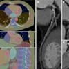
Lowering the radiation dose required for CT lung cancer screening exams has the potential to help allay concerns tied to radiation exposure. However, previous studies have reported inconsistencies in the categorization of lung nodules on very low-dose CT.
Tackling this issue, Marom and colleagues evaluated data from 36 patients who underwent both conventional low-dose CT and very low-dose CT at their institution. They applied their AI algorithm to the very low-dose CT scans, which allowed them to maintain the spatial consistency and fine detail of standard CT but at a markedly lower effective radiation dose (between 0.24 mSv and 0.41 mSv, compared with the 2 mSv of conventional CT).
The lung nodules found on the very low-dose CT scans with the AI algorithm received the same Lung Imaging Reporting and Data System (Lung-RADS) scores as those on the corresponding standard CT scans, unlike the assessment of very low-dose CT scans without the algorithm.
Lowering the radiation dose to less than 1 mSv for CT lung screening could lead to errors in the categorization of lung nodules, but implementing an AI algorithm could eliminate these errors, Marom told AuntMinnie.com.
"We showed that it is possible to screen patients for lung cancer with a radiation dose similar to two posterior-anterior and lateral chest radiographs using the artificial intelligence image denoising technology we developed," she said.