
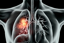
The International Association for the Study of Lung Cancer (IASLC) has emphasized again its support for CT lung cancer screening as a tool for identifying early-stage lung cancer and reducing the mortality rate of high-risk smokers.
Two large-scale randomized controlled studies -- the National Lung Screening Trial (NLST) and the Dutch-Belgian Randomized Lung Cancer Screening (NELSON) trial -- have demonstrated that lung screening can lead to significant mortality reduction in high-risk smokers. These results emphasize the need to provide routine early detection of lung cancer through screening as well as smoking cessation efforts, according to the IASLC.
Each national health service has unique challenges to consider when implementing CT lung screening programs. The IASLC pointed to several key practices that these programs should consider focusing on, including the following:
- Calling on a multidisciplinary group of experts
- Identifying high-risk individuals
- Acquiring consistent, high-quality CT scans
- Using defined clinical workup for indeterminate nodules
- Using an established process to manage suspicious nodules
- Integrating smoking cessation efforts into screening
"The unanimous consensus of the committee's screening experts is that now is the time for international leaders, governments, healthcare systems, and other stakeholders to implement global lung cancer screening programs, as they do for breast cancer (mammography) and colon cancer (colonoscopy), which save the lives of countless individuals," said Dr. James Mulshine, chair of the IASLC Early Detection and Screening Committee.


![Axial images from unenhanced calcium score cardiac CT (left) and curved planar reformation images from CT angiography (right) show that higher long-term exposure to air pollution is associated with greater coronary artery calcium and more obstructive coronary artery disease (CAD). Top row: Images in a 68-year-old male patient with higher 10-year mean ambient air pollution exposure (7.9 μg/m3 for particulate matter measuring ≤2.5 μm in diameter [PM2.5] and 17.4 parts per billion [ppb] for NO2) with extensive CAD (coronary artery calcium score [CACS] >1,000 and obstructive CAD [≥70% diameter stenosis]). Bottom row: Images in a 57-year-old female patient with lower 10-year mean ambient air pollution exposure (6.3 μg/m3 for PM2.5 and 4.6 ppb for NO2) with no CAD (CACS = 0 and no obstructive stenosis).](https://img.auntminnie.com/mindful/smg/workspaces/default/uploads/2026/06/hanneman.r6SMLzkezo.png?auto=format%2Ccompress&dpr=2&fit=crop&h=167&q=70&w=250)
