Outdated practices for cleaning CT scanners were identified as the cause of an ongoing hospital-acquired bacterial infection at the University of California, San Francisco (UCSF), according to a research letter published online October 23 in JAMA Internal Medicine.
Clostridium difficile is one of the most common causes of hospital-acquired infection, and it has been associated with significant morbidity and mortality, Dr. Sara Murray and colleagues wrote. Yet precisely where patients were catching the infection at UCSF was unknown.
In an effort to shed light on the matter, the researchers tracked patient movement in the hospital and its relation to the management of C. difficile infection by analyzing electronic health record (EHR) data between January 2013 and December 2015.
Among the nearly 87,000 hospitalized patients and 430,000 patient location changes, more than 1,100 cases involved C. difficile infection, Murray and colleagues found.
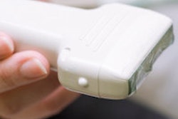
They discovered that exposure to a particular CT scanner in the emergency department was significantly associated with the development of C. difficile infection, with an incidence of 4% and an odds ratio of 2.5. The correlation was significant even after factoring in variables such as age, sex, number of location changes, length of stay, and prior hospitalization.
Upon further investigation, the researchers discovered the basis of the infection: The personnel responsible for cleaning that specific CT scanner table were not following updated cleaning procedures.
Identifying the root cause of the infection was just the first step in applying analytic methods to hospital data to instill real-world practice change, according to the researchers.
"Leveraging [electronic health record] data for spatial and temporal analytics may be a widely applicable strategy for infection control and quality improvement at other institutions and for other infectious diseases," they wrote.